GRANULOMATOUS INFLAMMATION
It is a form of chronic inflammation characterized by collections of “Activated” Macrophages, T lymphocytes and sometimes with necrosis.
Before we understand granuloma, let us understand the role of macrophages in inflammation.
Macrophages:
Are the dominant cells in most chronic inflammatory reactions.
These are derived from hematopoietic stem cells in the bone marrow in postnatal life. They are monocytes in the circulating system. The half life of these monocytes is one day! These monocytes differentiates into macrophages/histiocytes in the tissues. The half life of macrophages can vary from several months to years. These are diffusely scattered in various connective tissues.
However, there are macrophages with “special names” which are located in different locations. These are the ones which arise from progenitors in the yolk sac or fetal liver very early in embryogenesis. They migrate to the organs and persist for life
These are Kupffer cells, Sinus histiocytes, Microglial cells and alveolar macrophages in liver, spleen/lymphnodes, brain and lungs respectively as illustrated below.
Activation of Macrophages:
The monocytes in circulation reach the site of injury due to the presence of various chemotactic factors and differentiate into macrophages.
These macrophages have to be “activated” for them to be fully functional. The activation takes place either by classical(M1) or Alternative (M2)macrophage activation pathways as illustrated below.
The macrophages activated via classical activation are mainly “microbicidal” and “inflammatory”, whereas the macrophages activated via alternate activation are anti inflammatory in nature and help in tissue repair and fibrosis as illustrated below.
The main function of macrophages is to eradicate or eliminate the infective or offending agent. if they are unable to do so, then they try to “CONTAIN” the offending agent by forming a “Granuloma”. The granulomas function as a “rescue team” to contain the offending agent.
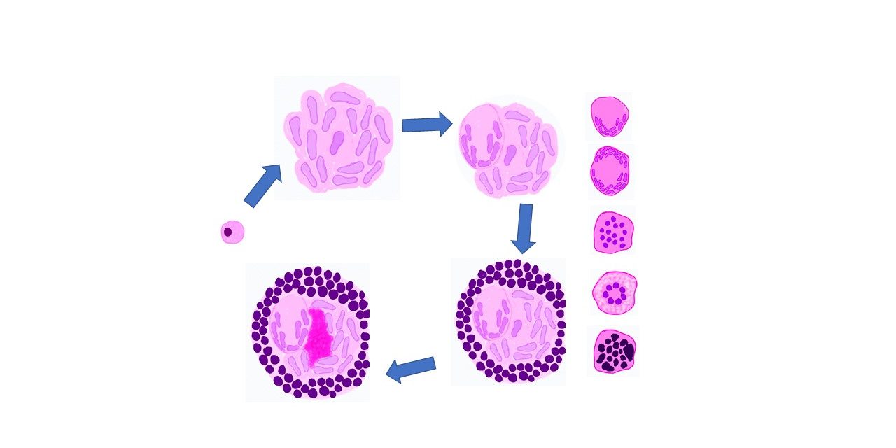
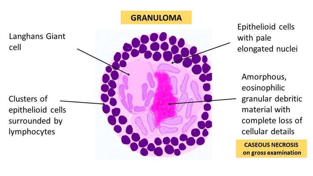
Tuberculosis is the prototype of granulomatous disease caused by infection. The activated macrophages in these granulomas are large, polygonal and have an oval/ elongated pale staining nuclei and they look like epithelial cells. Hence these cells are called as “Epithelioid cells”. Ultrastructurally, epitheliod cells also found to have “tight junctions”(like epithelial cells) and hence they can aggregate to form granulomas. The nucleus of these cells can be elongated and resemble the shape of sole/slipper and hence it is also referred to as “Slipper shaped” nuclei ( as described in some textbooks).
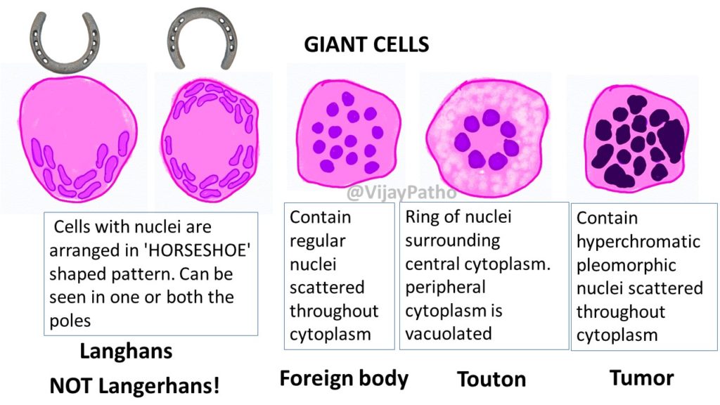
The epithelioid cells can fuse to form a multinucleated giant cells. in tuberculosis these are referred to as LANGHANS type of giant cells. These are large cells with abundant cytoplasm, and the nuclei are arranged in ‘HORSESHOE’ shaped pattern. The nuclei can be seen aggregated in one or both the poles of the cell.
These granulomas can be covered by a cuff of lymphocytes, plasma cells and macrophages. In long standing granulomas they can be surrounded by fibrosis
There can be necrosis in the center of these granulomas which on gross examination resembles that of a cheese and hence it is referred to as “CASEOUS NECROSIS”. Microscopically, it is amorphous, eosinophilic granular debritic material with complete loss of cellular details.
Examples of granulomatous inflammation
1. Tuberculosis
2. Leprosy
3. Syphilis
4. Cat scratch disease
5. Crohns disease, Sarcoidosis etc..
Different types of Giant cells
1. Langhans giant cells- seen in tuberculosis
2. Foreign body type of giant cells– formed as a reaction to insoluble exogenous or endogenous material, contain regular nuclei scattered throughout cytoplasm.
3. Touton giant cells- Giant cells with lipid filled(vacuolated) cytoplasm, seen in Xanthomas
4.Tumor giant cells– cells with numerous, hyperchromatic pleomorphic nuclei scattered throughout the cytoplasm, seen in most malignant tumors.
Other examples include Reed-Sternberg cells in Hodgkin lymphoma, Giant cells in Giant cell tumor/osteoclastoma, Aschoff giant cells in Rheumatic heart disease etc.
Recognition of granulomatous inflammation is very important as Tuberculosis is the prototype of a granulomatous disease caused by infection and should always be excluded as the cause when granulomas are identified.
CLICK HERE to read Pathology of Tuberculous Lymphadenitis
Watch Granulomatous inflammation on YouTube in the link below

