
Meningioma:
Meningioma is a predominantly benign tumour of meninges occuring in the adult population.
It arises from the meningothelial cells of the arachnoid.
These tumours are usually attached to the dura.
Meningiomas can also arise from the stromal arachnoid cells of the choroid plexus. In such instances, they can be found within the ventricular system or along any of the external surfaces of the brain.
Dural based masses – Differentials include
Meningiomas
Metastases
Solitary fibrous tumours
Poorly differentiated sarcomas
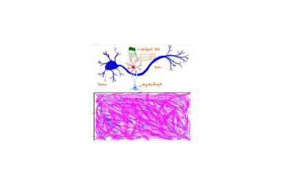
Pathogenesis of Meningioma: See the illustration below
Meningiomas are common in the setting of Neurofibromatosis 2(NF2). These harbour deletions in the 22q12 which encodes the gene NF2 and protein merlin.
In meningiomas not associated with NF2, the common mutations occur in TRAF7, KLF4, AKT1 and SMO.

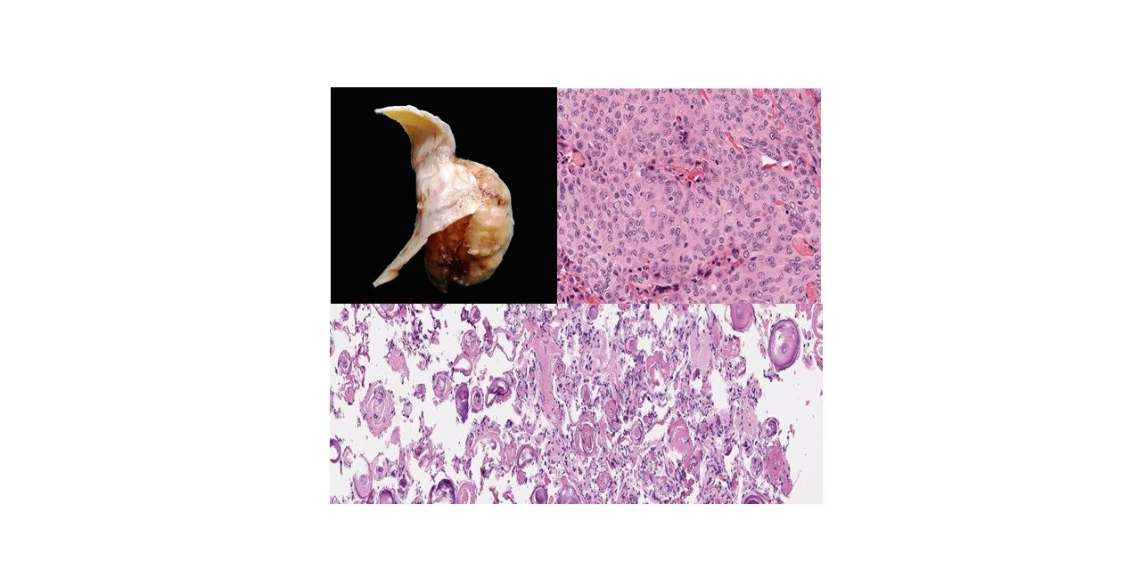
Morphology
On gross examinations, meningiomas are rounded, or bosselated masses with a rubbery consistency. They can be firm to finely gritty due to calcification.

Microscopy
Most of the meningiomas have a relatively low risk of recurrence and are WHO grade 1.
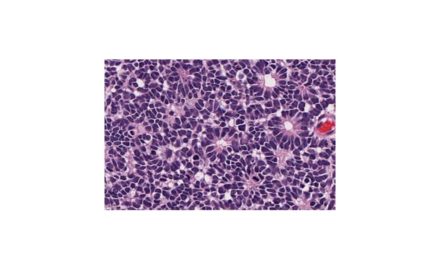
The tumour has a lobulated architecture.
Meningothelial cells are arranged in whorls, sheets, syncytium and nests.
These cells have eosinophilic cytoplasm, indistinct cell membrane and uniform nuclei. Nuclear grooves and intranuclear pseudoinclusions may be seen.
Psammoma bodies can be seen. Psammoma body – is a form of dystrophic calcification


Histologic variants of Meningioma
WHO grade 1: Low recurrence, Less aggressive
1. Meningothelial
2. Transitional
3. Fibroblastic
4. Psammomatous
5. Angiomatous
6. Secretory
7. Lymphocyte rich
8. Metaplastic
9. Microcystic
WHO grade 2: These have higher recurrence rates and more aggressive local growth
1. Atypical
2. Chordoid
3. Clear cell
WHO grade 3: Highly aggressive which mimics carcinoma/sarcoma.High recurrence with high mortality
1. Anaplastic
2. Papillary
3. Rhabdoid
Clinical features
Most of the meningiomas are slow-growing tumours. Patients present with vague symptoms or with focal findings related to the compression of the underlying brain parenchyma by the tumour.
Acknowledgements: Microphotographs -courtesy: pathpresenter.net
Note: The virtual slides of the annotated images can be accessed in the links below from Pathpresenter.net
Meningioma CLICK HERE
Psammomatous meningioma CLICK HERE