What is Warthin Tumor?
Warthin Tumor is a benign salivary gland tumor composed of oncocytic epithelial cells lining ductal, papillary, and cystic structures in a lymphoid stroma. It is the second most common salivary gland tumor after pleomorphic adenoma.
Why is it called Warthin Tumor?
The tumor is named after the American pathologist Alfred Scott Warthin, who made significant contributions to pathology. His name is also associated with:
Warthin-Finkeldey cells (seen in measles)
Warthin-Starry stain (used in diagnosing syphilis)
Warthin’s sign (accentuation of the pulmonary second sound, significant in pericarditis).
Is Warthin Tumor associated with smoking?
Yes, it is often referred to as the “smoker’s tumor” due to its strong association with smoking. Other risk factors include:
Radiation exposure (e.g., in atomic bomb survivors)
Autoimmune diseases, particularly thyroiditis.
What are the earlier terminologies for Warthin Tumor, and are they still used?
Previously, Warthin Tumor was called:
Adenolymphoma
Papillary cystadenoma lymphomatosum
Cystadenolymphoma
The World Health Organization (WHO) now recommends NOT using these terms.
What is the typical demographic affected by Warthin Tumor?
Commonly seen in males (more than females).
Occurs most often between the fifth and seventh decades of life.
Primarily arises in the parotid gland, with most cases being unifocal. However:
10% are multifocal.
10% are bilateral.
What is the etiopathogenesis of Warthin Tumor?
While the exact cause is not known, it is hypothesized to arise from salivary ductal inclusions in lymph nodes within the parotid gland.
What are the clinical features of Warthin Tumor?
Patients typically present with:
A painless, slow-growing mass in the parotid gland.
Occasionally, the mass may feel fluctuant on palpation.
A history of smoking often raises suspicion of Warthin Tumor.
How is Warthin Tumor diagnosed?
The initial diagnosis is often made using fine needle aspiration cytology (FNAC), which shows:
Sheets of oncocytes (large cells with abundant mitochondrial content and granular cytoplasm).
Numerous small lymphocytes.
Occasional larger lymphoid cells.
A granular and dirty background.
What are the gross features of Warthin Tumor?
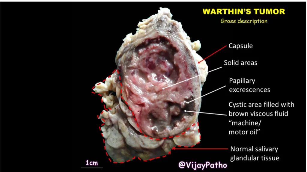
Encapsulated tumor with solid and cystic areas.
Papillary excrescences may protrude into the cystic spaces.
Cystic fluid is typically brown and viscous, often described as having a machine oil or motor oil appearance.
What are the microscopic features of Warthin Tumor?
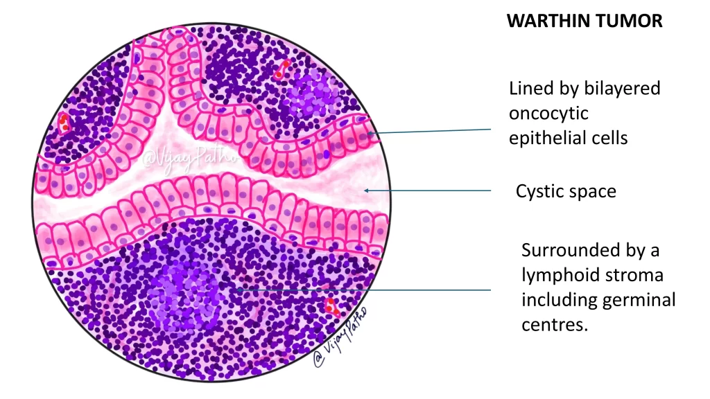
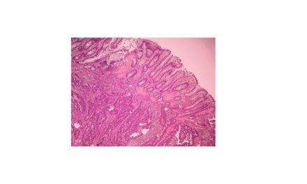
Papillary-cystic structures lined by bilayered oncocytic epithelial cells:
Outer cuboidal cells.
Inner columnar cells, facing the cystic spaces.
The epithelial lining is surrounded by lymphoid stroma, which may contain:
Prominent germinal centers.
Various proportions of lymphocytes and transformed lymphocytes.
The oncocytic cells appear granular under H&E staining due to their abundant mitochondria.
How is Warthin Tumor treated?
The primary treatment is complete surgical excision with adequate margins, which is typically curative.
Recurrence is rare.
CLICK BELOW TO WATCH THE VIDEO ON WARTHIN TUMOR

