
Pleomorphic adenomas are benign ‘mixed’ tumors that consist of varying proportions of epithelial and mesenchymal elements. The word “Pleomorphism” denotes the architectural complexity. They represent about 60% of tumors in the parotid and are associated with PLAG1 overexpression.
Pleomorphic adenomas mostly occur in the young- and middle-aged adults, between 30 and 60 years.
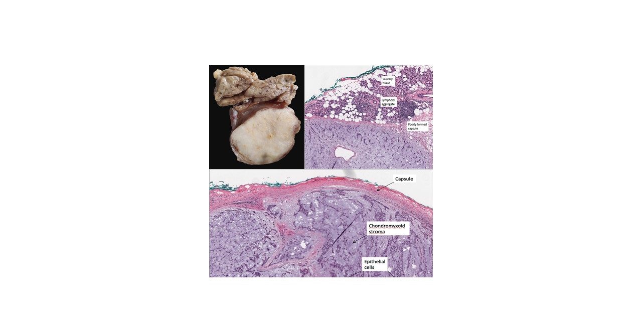
Gross Findings
The gross appearance is of a rounded, well-demarcated, glistening, expansile mass with poorly developed capsule. Microscopic protrusions into the surrounding salivary gland may lead to recurrences if the tumor is enucleated. Therefore, the treatment of choice is a parotidectomy.
 Photo credit: Vijay Shankar S
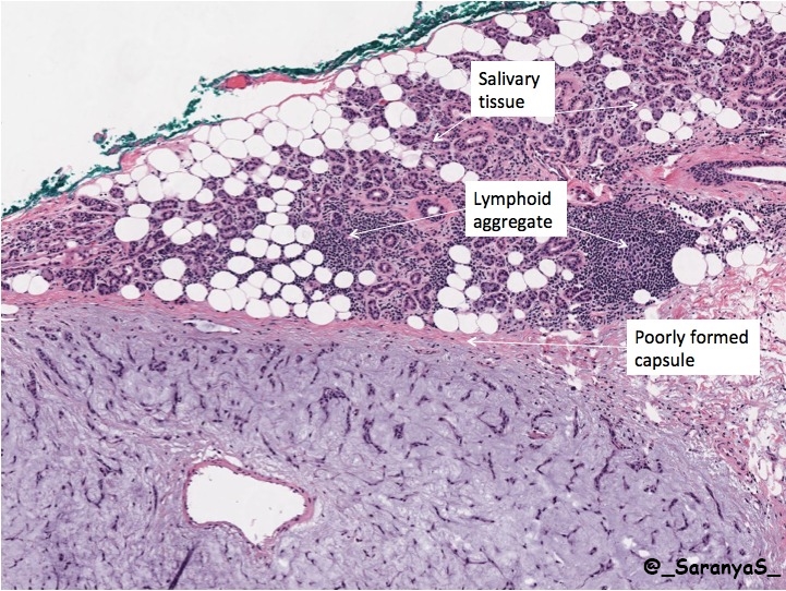
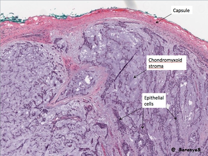
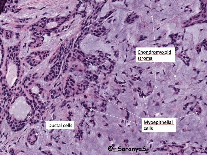
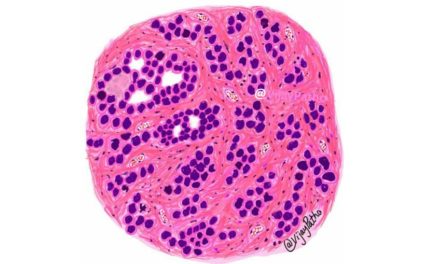
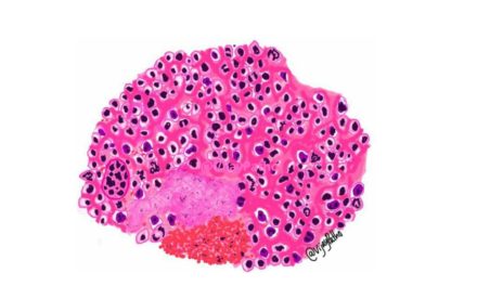
Microscopic Features
The dominant microscopic feature is tumor heterogeneity.
The epithelial elements resembling ductal cells or myoepithelial cells are arranged in duct formations, acini, irregular tubules, strands, or sheets of cells. The cells are dispersed in mesenchymal element consisting of a myxoid, hyaline, chondroid or osseous matrix.
A carcinoma that occurs in a pleomorphic adenoma is called a carcinoma ex pleomorphic adenoma or a malignant mixed tumor. They are highly aggressive and can take the form of an adenocarcinoma or undifferentiated carcinoma.
Microphotographs credit: Pathpresenter.net. The virtual slide of pleomorphic adenoma can be accessed below
CLICK HERE to view
