
MALIGNANT MELANOMA
Introduction
Melanoma or Malignant melanoma is the deadliest of all skin cancers.
It occurs commonly on the skin surface.
It can arise from oral mucosa, anogenital mucosa, meninges, uveal tissue, Gastro-intestinal tract, etc.
Though it has the suffix “oma”, melanoma is a malignant tumour
Risk factors
The risk factors for melanoma are:
Hereditary – can be inherited as autosomal dominant trait with variable penetrance.
Ultraviolet (UV) radiation exposure
Sunlight exposure
Fair skin
Severe sunburns
Etiopathogenesis
The driver mutations in melanoma involve the cell cycle control, pro-growth pathways and telomerase.
Some common mutations involving cell cycle control genes are CDKN2A which in turn encodes tumour suppressors like p15, p16 and ARF.
Mutations involving the pro-growth pathways include activating mutations in BRAF, loss of PTEN, loss of function mutation of NF1.
The common mutation involving telomerase is TERT (telomerase reverse transcriptase) gene.
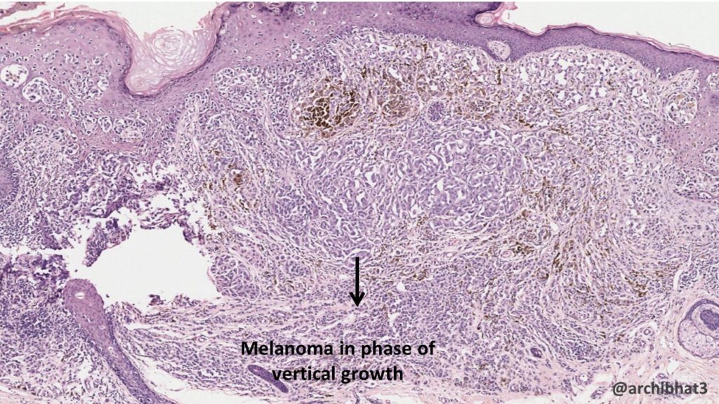
Progression of Melanoma
There are two growth phases in melanoma namely- the radial growth phase and the vertical growth phase.
In the phase of radial growth, the tumour cells spread horizontally in the epidermis and superficial dermis. In this initial phase, melanoma lacks the ability to metastasize.
The phase of vertical growth is characterised by downward growth of tumour cells into deeper dermis. This phase is often associated with the formation of nodule. The tumour acquires the ability to metastasize with the beginning of this phase.
Morphology
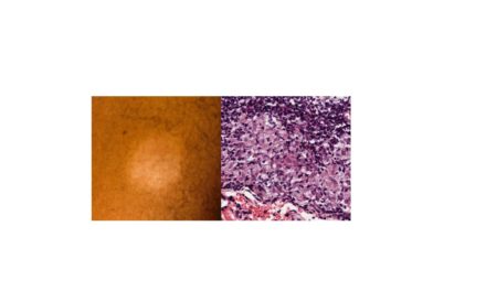
Melanomas show varying colours like black, brown, red, blue and gray.
Melanomas have irregular borders.
The benign counter-part or nevi are usually small, and show smooth and regular borders and uniform colour.
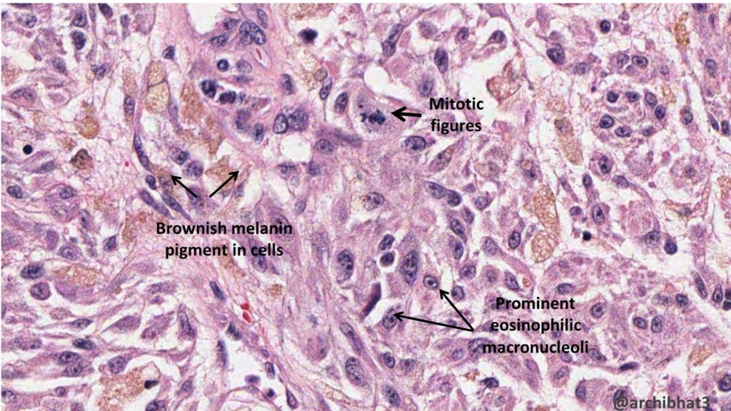
Microscopy
Individual melanoma cells have enlarged nuclei and large, prominent, eosinophilic nucleoli. The cells can be oval to round to spindle in shape.
Melanoma is called a great mimicker as the cells can show varied morphology.

The prognosis in melanoma depend on various factors like
1. Depth of invasion (Breslow thickness)
2. Mitotic count
3. Ulceration of overlying skin
4. Tumour infiltrating lymphocytes
5. Evidence of tumour regression
6. Location
Clinical features
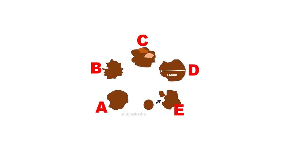
The warning signs of melanoma are called ADCDEs
A– Asymmetry
B- irregular borders
C- variegated colour
D- increasing diameter
E- evolution or rapid change

CLICK HERE to view virtual slide of superficial spreading melanoma from pathpresenter.net


