
Intradermal nevus
Melanocytes: these are the pigment producing cells in the skin which are present in the dermoepidermal junction. They are also seen in hair follicles.
Melanocytic nevi: benign tumors which are derived from melanocytes. They can be present at birth ( congenital melanocytic nevus) or may appear later in life ( acquired nevus)
Depending on the location of proliferating melanocytes in the skin, nevi can be one of the following three types
- Junctional nevus: melanocytes confined to epidermis only
- Intradermal nevus : melanocytes confined to dermis only
- Compound nevus : the nevi which have both epidermal and the derma component.
Clinical features: these are pigmented lesions of varying size ( 2- 6mm in diameter). The characteristic feature is that the lesion is uniform and has a symmetric architecture and appear as papule or macules.
Microscopy: The melanocytes in Junctional nevus appear as nests in the epidermis usually at the tips of rete ridges.
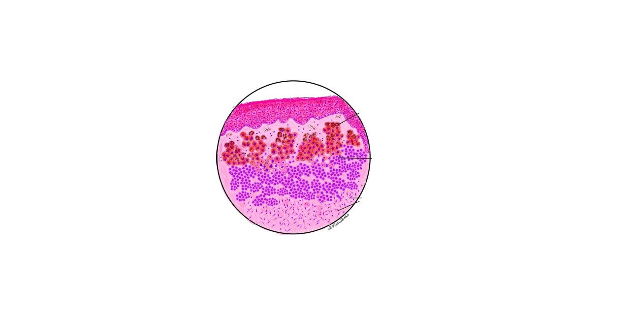
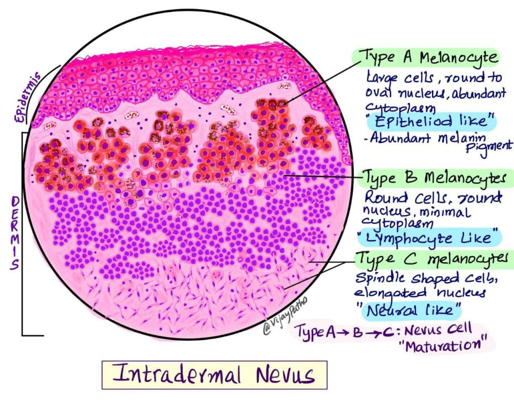
Microscopy of intradermal nevus: The tumor is symmetric and circumscribed. The melanocytes are predominantly arranged in nests and are monotonous in appearance with bland cytological features.
A very characteristic feature of intradermal nevus is its maturation. The cells show specific features as they progress deeper in the dermis. The most superficial location has larger nests, larger cells, more pigment and round to polygonal in shape. And all these changes as they progress below. Based on these features the melanocytes are divided into three types as below
- Type A melanocytes: these are larger cells, round to polygonal and have moderate to abundant cytoplasm with varying amounts of pigments in the cytoplasm, “Epithelioid- like”
- Type B melanocytes: the nests are smaller and the cells are smaller and round, “ Lymphocyte- like”.
- Type C melanocytes: These are spindle shaped cells with elongated nucleus and are found in the deeper dermis, “neural- like”. They do not form nests and are found individually.
