
What is Necroptosis?
Necroptosis is a type of cell death that appears like necrosis, but the mechanism behind it is programmed, similar to apoptosis.
So, you can think of it as a hybrid:
This is why necroptosis is also referred to as “programmed necrosis.”
How Does Necroptosis Resemble Necrosis Morphologically?
In necroptosis, the cell shows features commonly seen in necrosis:
-
Loss of ATP
-
Swelling of the cell and its organelles
-
Release of lysosomal enzymes
-
Reactive oxygen species generation
-
Rupture of the plasma membrane
Once the membrane ruptures, the cell contents spill out, triggering inflammation and tissue damage, just like in necrosis.
How is Necroptosis Different from Apoptosis?
The key difference is in the molecular pathway:
| Apoptosis | Necroptosis |
|---|---|
| Controlled cell death | Controlled cell death |
| Caspase dependent | Caspase independent |
| Does not cause inflammation | Causes inflammation |
In necroptosis, caspases (especially caspase-8) are either not present or inhibited.
Which Key Proteins Are Involved in Necroptosis?
Necroptosis involves specific kinases:
-
RIPK1 (Receptor Interacting Protein Kinase 1)
-
RIPK3 (Receptor Interacting Protein Kinase 3)
These two come together to form a complex that activates another protein called MLK
(you mentioned it as Mixed Lineage Kinase domain-like protein).
When MLK converts from a monomer to polymers, it moves to the cell membrane and creates pores.
These pores allow ions to rush in → osmotic swelling → membrane rupture → inflammation.
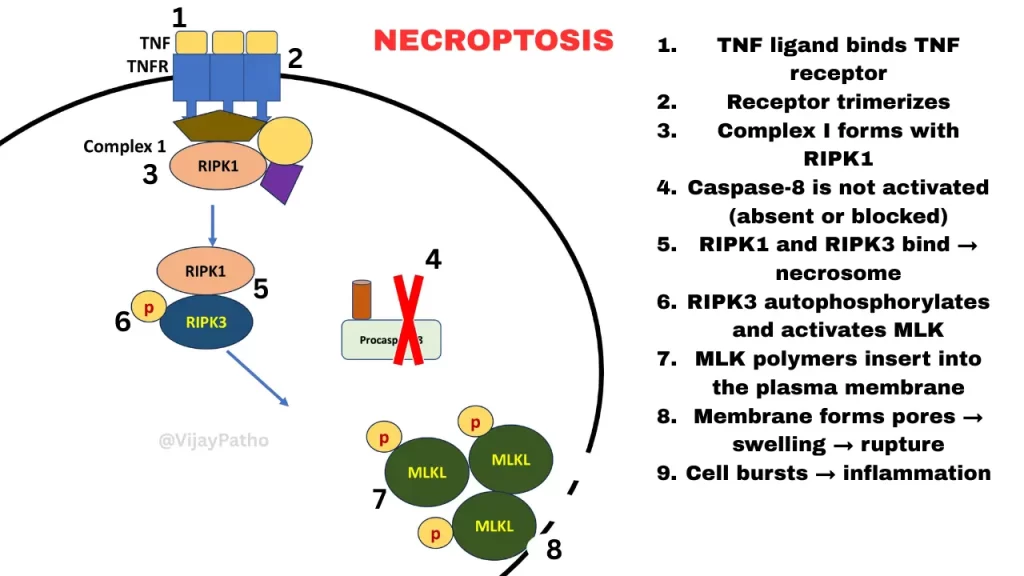
Steps of Necroptosis (Simplified)
-
TNF ligand binds TNF receptor
-
Receptor trimerizes
-
Complex I forms with RIPK1
-
Caspase-8 is not activated (absent or blocked)
-
RIPK1 and RIPK3 bind → necrosome
-
RIPK3 autophosphorylates and activates MLK
-
MLK polymers insert into the plasma membrane
-
Membrane forms pores → swelling → rupture
-
Cell bursts → inflammation
Where Does Necroptosis Occur in Normal Physiology?
Bone growth plate formation in mammals.
Chondrocytes undergo necroptosis during ossification.
Where Is Necroptosis Seen in Disease?
Some important pathological examples:
-
Steatohepatitis (liver cell death)
-
Acute pancreatitis (death of pancreatic acinar cells)
-
Ischemia-reperfusion injury (brain, heart, kidney)
-
Neurodegenerative disorders like Parkinson’s disease
Role of Necroptosis in Host Defense
Some viruses produce caspase inhibitors to block apoptosis and keep the host cell alive longer.
When apoptosis is blocked, necroptosis acts as a backup, killing the virus-infected cell and preventing further viral replication.
So necroptosis helps in limiting viral spread.
In Summary
Necroptosis is:
-
Morphologically similar to necrosis
-
Mechanistically programmed like apoptosis
-
Caspase-independent
-
Mediated mainly by RIPK1, RIPK3, and MLK
-
Leads to inflammation
-
Plays roles in normal development, disease, and viral defense
CLICK below to view the video tutorial on Necroptosis
