
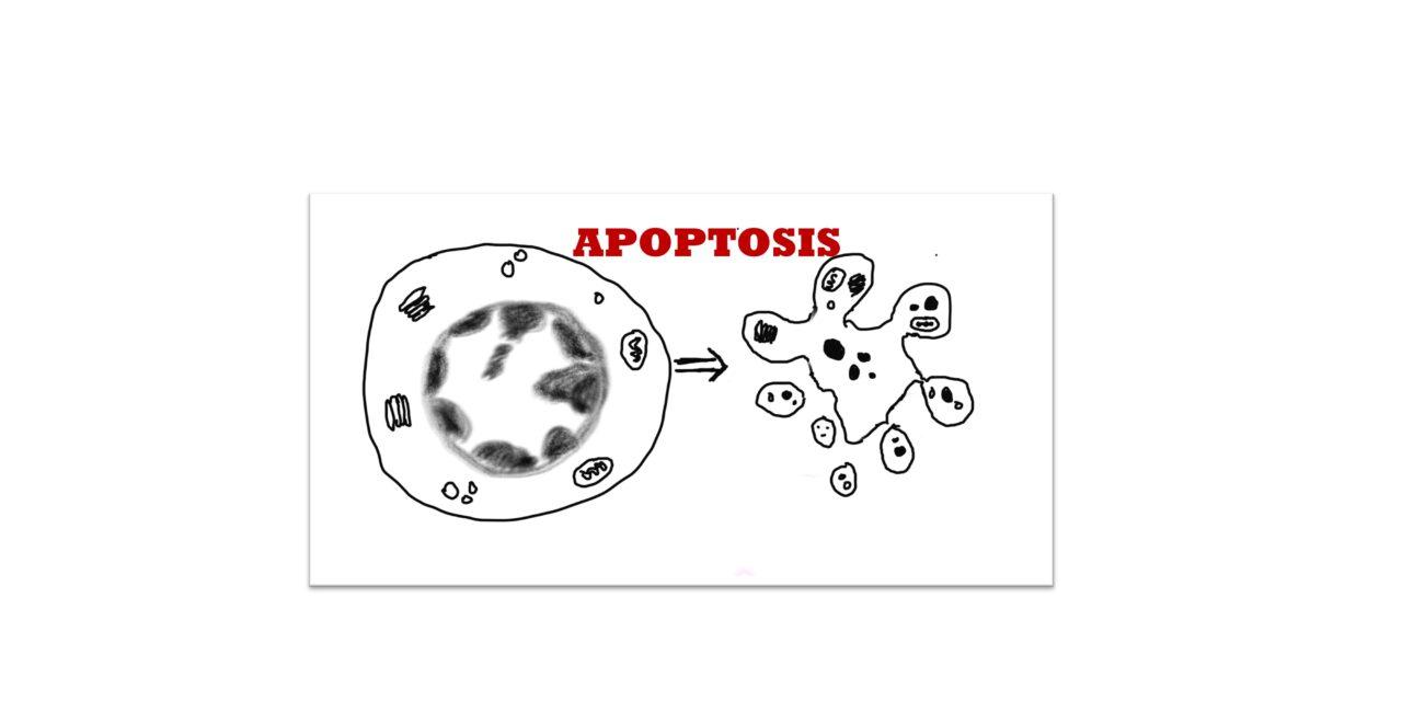
Apoptosis (pronounced apo- toe-sis)
1 Introduction
Cells can die in many ways and for many reasons
There are several pathways of cell death (more than 20 are identified at present) among which the ones which have been most studied are necrosis (also called accidental cell death) and apoptosis (also called programmed cell death)
During apoptosis the cell moves through a set of activities in a coordinated manner to achieve the end result i.e., cell death. Hence it is termed as ‘programmed’
2 Is apoptosis good or bad?
Apoptosis is essential during organogenesis/ formation of the body in utero to give correct structure to various organs like making individual fingers of the hand by removing the webs from between the fingers.
Apoptosis is also a mechanism of aged cells or irreversibly damaged cells to die
Apoptosis is one of the mechanisms how the cancerous cells, microorganisms and self-reactive lymphocytes (lymphocytes that cause autoimmune disease) are made to die by our immune system.
Apoptosis causes several neurodegenerative diseases like Huntington’s disease and Parkinsons disease.
Hence apoptosis is neither good nor bad. Its just where and when it happens determines whether its beneficial or harmful for the body
3 Why do you need to know apoptosis?
Because it’s an exam question
Because many physiological and pathological process use apoptosis in their mechanism and it’s important to understand it.
Currently there are drugs which are targeting the mechanism of apoptosis for treatments and understanding apoptosis gives one a better understanding of their usage.
4 Apoptosis following cell injury
Cells injured in many ways (refer causes of cell injury) can choose apoptosis as mechanism of death. However classically apoptosis follows damage by irradiation and chemotherapeutic drugs which irreversibly damage the DNA. Other causes include absence of growth factors and misfolded protein accumulation.
5 Mechanism ( Fig 1)
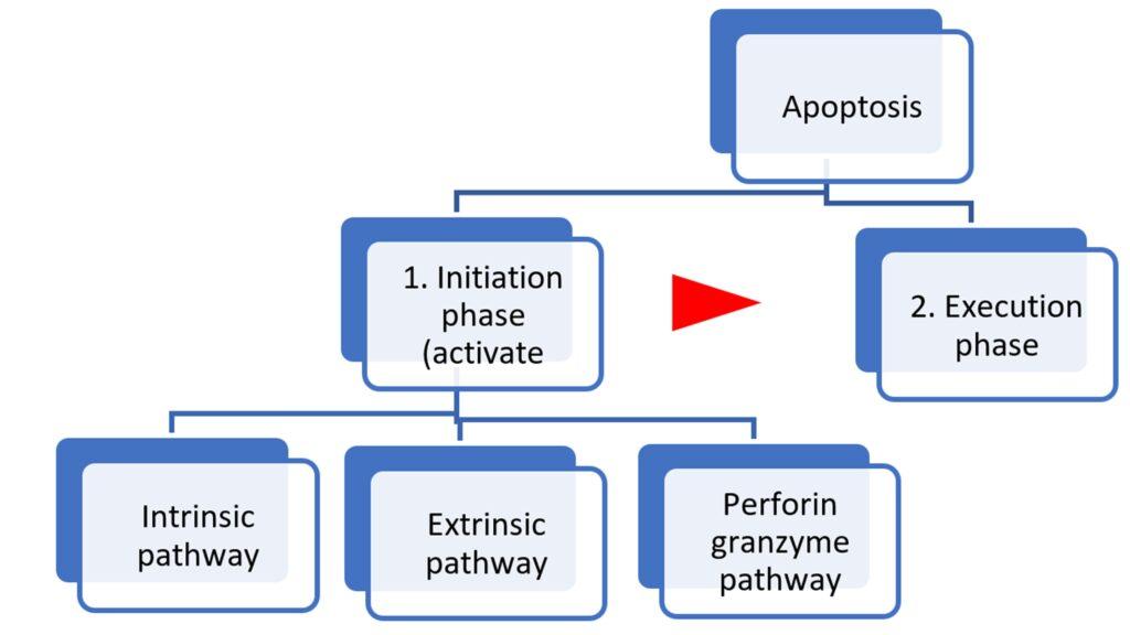
There are two phases – I Initiation phase II execution phase
Apoptosis is mainly brought about by two sets of proteases called Caspases -the initiator caspases ( 8.9 and 10) and the effector caspases (3,6,7) which take part in the initiation and execution phases respectively.
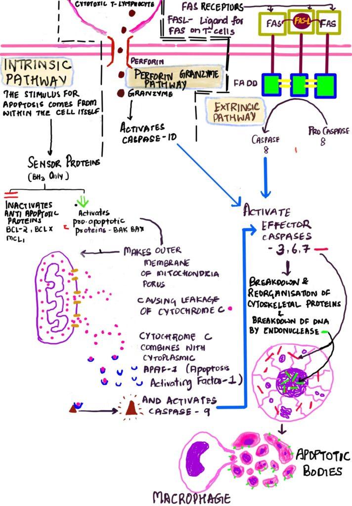
Fig : 1 Graphic representation of two phases of apoptosis.
5.1 Initiation of Apoptosis
Can occur by three pathways– intrinsic pathway, extrinsic pathway, and the perforin granzyme pathway
5.1.1 Intrinsic pathway –
The signal for apoptosis comes from within the cell hence the term intrinsic
This pathway is regulated by two sets of proteins as illustrated below
Balance is maintained by sensor proteins
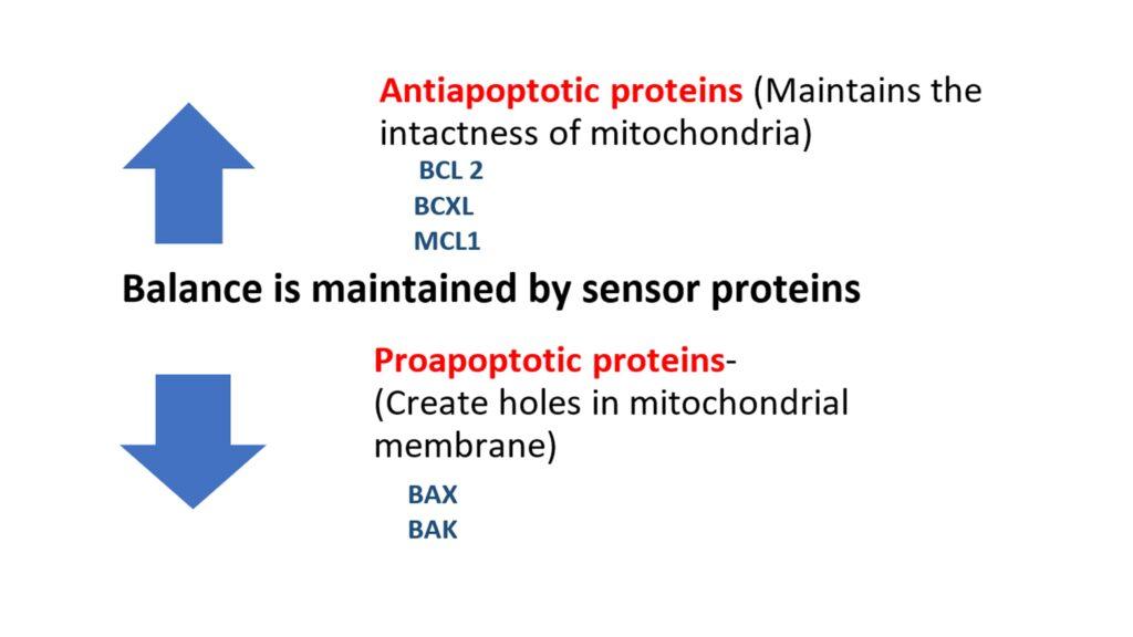
Any stimulus that informs the sensor proteins to initiate apoptosis will result in increase in the proapoptotic set. The BAX and BAK, once activated, form pores or channels in the outer mitochondrial membrane, leading to the release of Cytochrome c from the mitochondrial intermembrane space. Cytochrome C, once released into the cytoplasm, binds to Apaf-1 (apoptotic protease-activating factor 1) forming a complex called the apoptosome.
This complex activates caspase-9, an initiator caspase. Activated caspase-9 then cleaves and activates effector caspases, such as caspase-3 and caspase-7, which are responsible for the execution phase of apoptosis
5.1.2 Extrinsic pathway- the signals come from outside the cell hence the term extrinsic
The cells in the body have a receptor on their surface called the death receptor. FAS is one type of death receptor. It gets activated when its ligand (FASL) binds it. FASL is found on the surface of T lymphocytes. Once the ligand binds, the receptors make themselves into a set of three(trimerization).
Within the cell the FAS receptor has another protein linked to it called Fas Associated Death Domain( FADD). Once the FAS receptors trimerise the FADD forms a complex with procaspase-8 (another initiator caspase) to form a complex known as the Death-Inducing Signaling Complex (DISC) that activates the procaspase 8 to Caspase 8 which then activates the effector caspases 3,6 and 7
5.1.3 Perforin-mediated cell lysis
This is done by the cytotoxic T Cells which use this pathway to kill cells containing microbes. The CTLs recognize abnormal or infected cells through the presence of specific antigens on the cell surface when presented along with class I MHC molecules. They then release perforin, which forms pores in the target cell’s membrane. The pores created by perforin allow granzymes, into the target cell. Granzymes, once inside the target cell, activate caspases 10 and finally caspase 3 and initiate apoptosis.
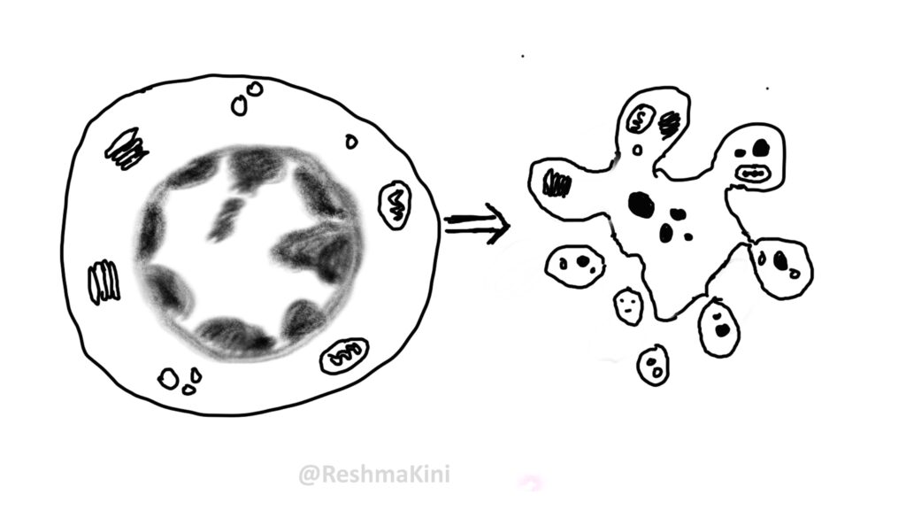
5.2 Execution phase
The execution phase of apoptosis involves the activation of effector caspases- 3,6,7.These cause breakdown of cytoskeletal proteins and activation of endonucleases which break the DNA into lengths of 180-200 base pairs.
Execution phase results in the cells breaking down into small membrane bound parts called apoptotic bodies. These apoptotic bodies flip the tasty phosphatidylserine from the inner surface of their cell membrane to the outer surface. This tasty bit is easily recognized by the local macrophages and the apoptotic bodies get phagocytosed by them . Other chemicals that can make the apoptotic bodies tastier include thrombospondin, antibodies and complement C1q. Because the apoptotic bodies get consumed by the macrophages very fast, they do not release their content into the surrounding environment . Hence there is no inflammation following cell death by apoptosis. ( unlike in necrosis)
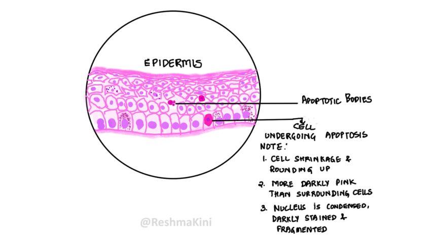
6 Morphology
Characteristic features are
1. Cell shrinkage
2. Condensation of nucleus- karyopyknosis followed by karyorrhexis
3. Formation of apoptotic bodies
Light microscopy
1. The cells are smaller, rounded and the cytoplasm is more darkly pink
2. The nucleus initially appears smaller and darker but eventually is fragmented
3. Apoptotic bodies appear as small intensely stained small rounded structures.
Electron microscopy
1. Cell shrinkage and the organelles get tightly packed
2. Condensation of chromatin under the nuclear membrane followed by breakup of the nucleus
3. Formation of cytoplasmic blebs. These blebs eventually get pinched off; the plasma membrane seals itself around the pinched off portions to form apoptotic bodies.
Summary
Apoptosis is one of the ways in which cell death occurs.
The mechanism is highly organized and hence its termed programmed cell death.
Caspases are endoproteases that are responsible for both initiation and execution of apoptosis
Initiation phase of apoptosis can occur by the signals that come from within the cell( intrinsic ) or from outside the cell ( extrinsic). It results in activation of the initiator caspases – 8,9,10.
Irrespective of how its initiated it follows a common execution pathway involving the effector caspases- 3,6,7
Effector caspases along with endonuclease breakdown the cytoskeleton and DNA of the cell.
Once the skeleton is broken the cell membrane undergoes blebbing and formation of apoptotic bodies which make themselves tasty to the macrophages. Apoptotic bodies get eaten by the macrophages. Since the cells don’t release their content in the surrounding environment there is no inflammation.
References
Elmore S. Apoptosis: a review of programmed cell death. Toxicol Pathol. 2007 Jun;35(4):495-516. doi: 10.1080/01926230701320337.
Saraste, A., & Pulkki, K. (2000). Morphologic and biochemical hallmarks of apoptosis. Cardiovascular research, 45(3), 528–537. https://doi.org/10.1016/s0008-6363(99)00384-3
Robbins and Cotran -Pathologic Basis of Disease 9th Edition
CLICK BELOW to view video tutorial