
Understanding Aneurysms: Types, Causes, and Management
What is an Aneurysm?
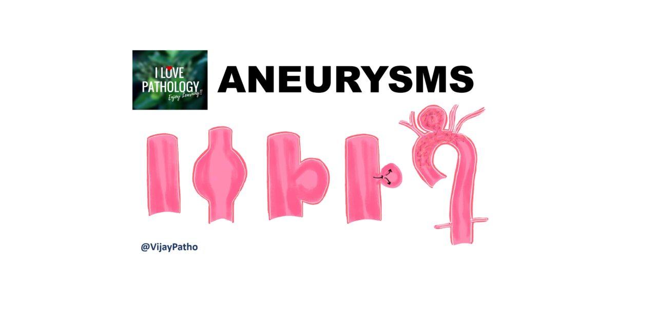
An aneurysm is a localized abnormal dilatation of a blood vessel or the heart, which can be congenital or acquired. To illustrate, picture a normal blood vessel with a uniform caliber. In contrast, an aneurysm represents an abnormal dilation of the blood vessel, as shown below:

Aneurysms can be classified as true aneurysms and false aneurysms (also known as pseudoaneurysms).
True Aneurysm vs. False Aneurysm
True aneurysms involve all the layers of an intact arterial wall or the thinned ventricular wall of the heart. Examples of true aneurysms include atherosclerotic and congenital vascular aneurysms, as well as ventricular aneurysms, particularly post-transmural myocardial infarction.
False aneurysms (pseudoaneurysms), on the other hand, represent a defect in the vascular wall leading to an extravascular hematoma that freely communicates with the intravascular space. Unlike true aneurysms, false aneurysms do not involve all the layers of the blood vessel. Examples include ventricular rupture after myocardial infarction and leaks at the suture junction of a vascular graft with a natural artery.
Both true and false aneurysms can rupture, leading to catastrophic consequences.

Macroscopic Classification
Macroscopically, aneurysms can be categorized based on their shape. The shape classification includes saccular aneurysms, which feature a spherical outpouching involving a portion of the vessel wall, and fusiform aneurysms, which involve diffusely circumferential dilatation of a long vascular segment.

Mechanisms of Aneurysms
Based on the mechanisms of aneurysm formation, they can be categorized as:
- Atherosclerotic aneurysms
- Syphilitic aneurysms
- Mycotic aneurysms (resulting from infections)
- Dissecting aneurysms, where blood enters a separated or dissected wall within the vessel.
The pathogenesis of aneurysms involves a compromise in the synthesis, degradation, and repair of the extracellular matrix, which maintains the structural and functional integrity of blood vessels. Various defects, such as poor-quality vascular wall connective tissue, abnormal transforming growth factor beta signalling, imbalance in collagen degradation and synthesis due to inflammation, and loss of smooth muscle cells or inappropriate synthesis of non-collagenous or non-elastic extracellular matrix, can lead to aneurysm formation.
Other risk factors contributing to vessel wall weakness include advanced-stage smoking, trauma, vasculitis, congenital defects, and infections.
Aortic Aneurysms
Now, let’s understand aortic aneurysms, which can occur in different parts of the aorta: the ascending aorta, the arch of the aorta, and the descending aorta (which is further divided into thoracic and abdominal aorta).
Abdominal Aortic Aneurysms
Abdominal aortic aneurysms are more common in men and smokers and are typically located between the renal arteries and the bifurcation of the aorta. They can be either circular or fusiform and are often larger than three cm in diameter, sometimes reaching up to 25 cm
Variants of Abdominal Aortic Aneurysms
- Inflammatory abdominal aortic aneurysms account for around 5-10% of cases and are characterized by abundant lymphoplasmacytic inflammation.
- Immunoglobulin G4-related disease can also affect abdominal aortic aneurysms, presenting with storiform fibrosis and high levels of circulating immunoglobulin G4.
- Mycotic abdominal aortic aneurysms are infected lesions that are prone to destruction of the vessel wall.
Clinical Manifestations and Management
Clinical manifestations of abdominal aortic aneurysms often include being asymptomatic or presenting as a pulsating abdominal mass. However, aneurysms can lead to catastrophic consequences such as rupture, obstruction of branch vessels resulting in tissue ischemia, embolism, and compression effects.
Management of abdominal aortic aneurysms involves aggressive approaches for aneurysms larger than five centimeters, including open surgery with prosthetic grafts or endovascular stent grafts. Early intervention is crucial, as the operative mortality is much higher if an aneurysm ruptures.
Thoracic Aortic Aneurysms
Thoracic aortic aneurysms are commonly associated with hypertension and other conditions such as Marfan syndrome, Loeys-Dietz syndrome, and aortitis. While most cases are asymptomatic, compression or catastrophic events can cause symptoms.
Syphilitic (Leutic) Aneurysms
Syphilitic aneurysms, also known as leutic aneurysms, are seen in the tertiary stage of syphilis. They are more common in men and often affect the ascending aorta and aortic arch. The pathogenesis involves inflammation around the vasavasorum of the adventitia, resulting in endarteritis obliterans and weakening of the vessel wall. Intimal surface is wrinkled, giving the classical TREE-BARK APPEARANCE

Conclusion
In conclusion, aneurysms are abnormal dilations of blood vessels or the heart that can lead to severe complications if not managed appropriately. Understanding their types, causes, and manifestations is essential for early detection and intervention.
Click here to view the Video tutorial on Aneurysm
