

Dysgerminoma -ovary
This is the ovarian counterpart of seminoma in males.
Gross: mostly unilateral, can be of variable size. This is encapsulated with smooth or bossellated external surface. Cut surface will be fleshy and solid, which is greywhite to yellowish.
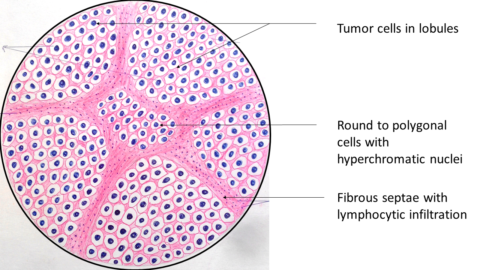
Microscopy: the tumor cells are arranged in sheets, groups which are separated by fibrous stroma.
The tumor cells are large and have clear cytoplasm, with well defined boundaries. The nucleus is centrally placed and is hyperchromatic, regular with prominent nucleoli.
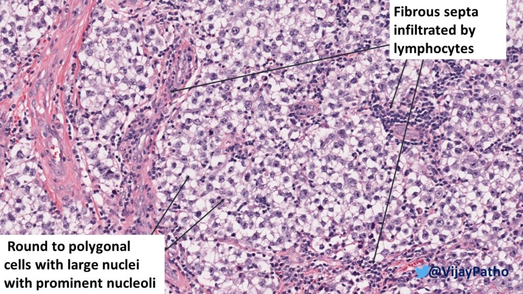
The fibrous stroma is infiltrated by lymphocytes which may be slight to marked. Sometimes lymphoid follicles are also seen. (it is proved that more lymphocytic infiltration is associated with better prognosis of these tumors!)
Rarely granulomas can also be seen.
The virtual slide of dysgerminoma can be accessed in the link https://www.pathpresenter.net/publicDisplay/DisplayCase/9962d7da-e932-4971-9254-1562517abc68#
Visit pathpresenter.net for amazing collection of slides for learning.

