

Leiomyoma -uterus
Benign smooth muscle neoplasm of uterus
Gross: circumscribed, mass of varying size with greywhite and whorled cut surface. They can be single or multiple. If the tumor is very large, the degenerative changes can be seen. Based on the location of the tumor they are categorized as
- Submucosal: present benath the endometrium. These are the ones which can manifest with menorrhagia
- Intramural: within the myometrium. These are usually asymptomatic if small and are often incidental finding.
- Subserosal / pedunculated: they are seen beneath the serosa. Can cause pressure symptoms due to compression on adjoining structures.
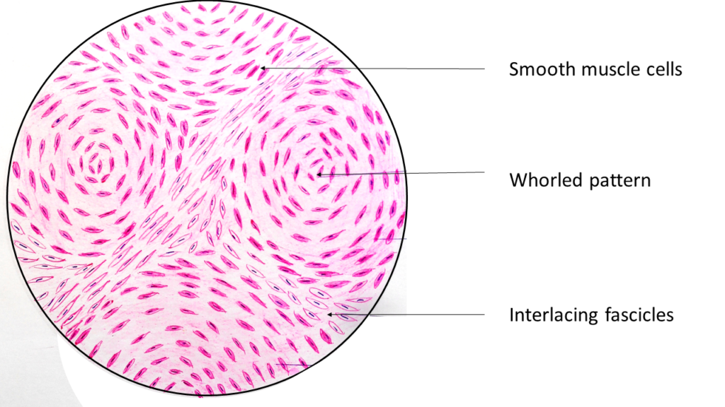
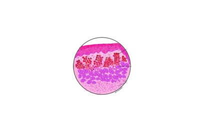
Microscopy: the pattern is that of interlacing fascicles or whorling of smooth muscle cells. The individual smooth muscle cells are uniform in size and shape and have characteristic oval to elongated with blunt ends ( cigar shaped). The cytoplasm s abundant and eosinophilic with elongated bipolar processes.
The leiomyomas are prone for development of secondary changes like
- Hyaline change
- Mucoid change
- Cystic change
- Calcification
- Red degeneration: seen in pregnancy and in oral contraceptive use. It is due to extensive coagulative necrosis and grossly it appears as homogeneous dark red.


Recent Comments