
- What is a Giant cell tumor of bone.
Giant cell tumor is a locally aggressive neoplasm of uncertain origin which affects predominantly the epiphysis of long bones. It is so called because the tumor is predominated by the presence of multinucleated osteoclast like giant cells. It was also referred to as osteoclastoma for the same reason. Very rarely they can involve metaphysis as well as joints.
- Discuss the epidemiology of Giant cell tumor of bone
Giant cell tumor commonly affects young adults, in age group of 20 – 40 years, with a slight female preponderance. Majority of the cases affect the distal femur or proximal tibia.
- What is the pathogenesis of Giant cell tumor of bone
Based on the experimental evidence, it is suggested that the osteoblast precursors, which are the minority in the tumor, are actually the neoplastic cells. These cells express Receptor activator of nuclear factor kappa-B ligand ( RANKL). RANKL results in recruitment of monocyte precursors and also promotes the formation of osteoclast like giant cells.
- What is the characteristic radiological appearance of Giant cell tumor of bone
Radiologically, these tumors appears large, eccentric lobulated lytic lesions. The lesion, is seen with or without trabeculations, and if trabeculations are seen, it gives the classic “soap bubble” appearance.
Soap bubble appearance is basically due to multiloculation and trabeculations.
- What are the clinical features of Giant cell tumor of bone
The patients present with swelling and pain in the joints specially on weight bearing and on movement .Pathological fractures can be rarely seen. On examination, the swelling will usually eb soft in consistency with a feeling of “egg shell crackling”.
- What are the 4 “E’s of Giant cell tumor?
These describe the features of Giant cell tumor in the form of Mnemonics!
Epiphyseal
Eccentric
Expansile
Egg Shell crackling
- Describe the gross appearance of Giant cell tumor of bone
These are usually large tumors, which are ephyseal and eccentrically located at the ends of long bone. The tumor is usually well circumscribed and dark tan in appearance. On cut section, the tumor appears haemorrhagic/necrotic with cystic areas.
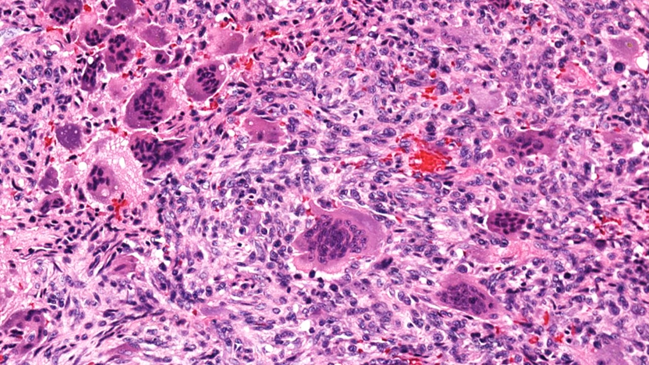
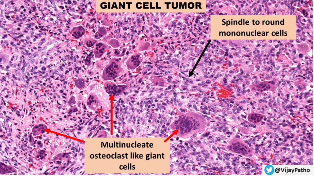
- Describe the microscopic appearance of Giant cell tumor of bone
The characteristic feature is the presence of numerous multinucleate osteoclast like giant cells scattered throughout the mononuclear cells in the stroma.
a. The mononuclear cells are the tumor cells. These are uniform cells, plump spindle to round to oval cells with the presence of large nuclei with prominent nucleoli. The cytoplasm if scanty.
The biological behavior of the tumor is determined by the histologic appearance of these tumor cells. Mitotic activity can be seen.
b. Multinucleated giant cells: these are osteoclast like giant cells with more than 100 nuclei in many of these cells. Note that the nuclei of these giant cells, resemble that of the nuclei of mononuclear stromal cells.
Stroma may show cystic changes in the form of presence of macrophages. Areas of hemorrhage and necrosis can be seen.
- Mention the other bone lesions which contain giant cells/ Mention the giant cell rich lesions of bone.
Multinucleate giant cells are seen in conditions other than giant cell tumors. These include
Benign conditions
- Chondromyxoid fibroma
- Chondroblastoma
- Non ossifying fibroma
- Aneurysmal bone cyst
- Osteoblastoma
Malignant lesions
- Osteosarcoma
- Clear cell chondrosarcoma
- Metastatic carcinomas
