Rhinoscleroma is an infectious chronic granulomatous disease of the upper respiratory tract caused by Klebsiella. rhinoscleromatis.
Klebsiella rhinoscleromatis is an immotile, short, encapsulated, Gram-negative bacillus which has an affinity for nasal mucosa.
Rhinoscleroma is transmitted by direct or indirect contact with nasal exudate of infected person. The most common site of involvement is the nasal cavity followed by nasopharynx, larynx , trachea and bronchi.
Clinical features: They present in 3 different stages
- Catarrhal-atrophic stage– associated with recurrent sinusitis and rhinorrhea
- Granulomatous-hypertrophic stage– associated with mass forming lesion with signs of obstruction.
- Sclerotic phase– chronic with extensive scarring and fibrosis.
Microscopic features:
Depends on the stage of the disease.
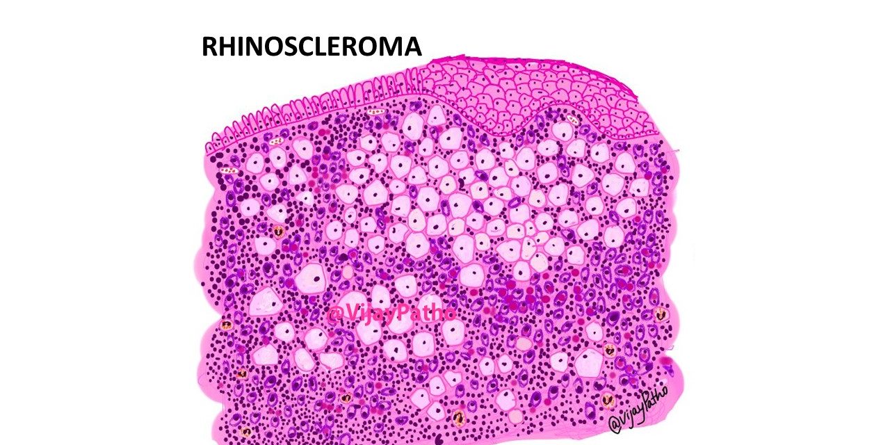
In the catarrhal stage non specific acute inflammation along with squamous metaplastic changes are seen.
The classical morphology with presence of MIKULICZ cells are observed in granulomatous hyperplastic stage. These cells are large , foamy histiocytes which when stained with gram stain, show numerous gram – negative bacilli within them.
These cells are surrounded by dense chronic inflammatory cell infiltrates comprised of plasma cells and lymphocytes. Numerous RUSSEL bodies are also noted with the plasma cells and in the stroma. Neutrophils and eosinophils can also be seen.
Russel bodies are large,eosinophilic, homogeneous immunoglobulin-containing inclusions. They are seen mainly within the plasma cells but can be seen extracellularly as well. They represent aggregates of immunoglobulin components which are not released due to a probable block in the normal secretion pathway of immunoglobulin.
In the sclerotic stage, extensive fibrosis , chronic inflammation is seen with few or absent Mikulicz cells.
Treatment: combination of antibiotics and surgical debridement. Recurrence is common.


