
Rhinospoiridiosis
It is a chronic granulomatous disease caused by Rhinosporidium seeberi which belong to class Mesomycetozoea .
Mesomycetozoea, are new group of microorganisms which lie in between fungi and animals and are usually parasites of fish and other animals.
This disease is endemic in India, Srilanka and Africa.
Mode of transmission: 1. By bathing in stagnant ponds in which animals also bathe;
2. Autoinoculation – rare
3. Hematogenous spread- rare
Clinical features: depends on the site of involvement. The lesions are usually seen in nasal cavity and nasopharynx. These are reddish polypoidal, bulky, friable mucosal masses. conjunctiva, mouth, larynx, genitalia and skin are the other rare sites of involvement.
In the nasal cavity the lesions begin as sessile masses and they gradually grow to become fleshy pedunculated polypoid mass.
Gross: These are reddish polypoidal, bulky, friable mucosal masses.
Microscopy: Has characteristic morphology.
Sporangia of various sizes are located in the stroma of the polyp. The overlying mucosa may show squamous metaplastic changes. The largest of these are usually immediate subepithelial in location which may or may not show evidence of rupture.
 Photo Credit: Dr Saranya Singaravel
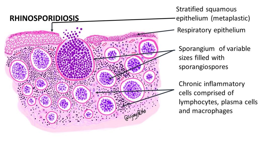
Mature sporangia are 100 to 450 micrometer in diameter with a thick chitinous wall and contain sporangospores in different stages of development. sporangiospores stain basophilic and measures 7 to 9 microns in diameter.
If there is rupture of sporangia in the stroma, an intense granulomatous response can be seen.

The stroma is comprised of moderate to abundant chronic inflammatory cell infiltrates made of lymphocytes, plasma cells and macrophages.
Treatment: Excision of the lesion is the treatment of choice

