
Inflammatory Dermatoses Reaction Patterns
This blog is designed for residents and consultants in pathology to provide a structured approach to interpreting inflammatory dermatoses, which will be especially helpful during routine reporting.
Inflammatory dermatoses encompass a wide range of skin conditions characterized by distinct patterns of inflammation. Identifying these reaction patterns is essential for pathologists to make accurate diagnoses, as different dermatoses can exhibit overlapping features.
By adopting an algorithmic approach, one can systematically evaluate histopathological features to narrow down the differential diagnosis.
The six main reaction patterns in inflammatory dermatoses are:
Lichenoid Reaction Pattern – Often involves inflammation at the dermoepidermal junction, with characteristic changes in the basal layer.
Psoriasiform Reaction Pattern – Characterized by epidermal hyperplasia with varying degrees of parakeratosis and acanthosis.
Spongiotic Reaction Pattern – Marked by intercellular edema within the epidermis, commonly seen in eczematous conditions.
Vesiculobullous Reaction Pattern – Defined by the formation of blisters at different epidermal levels, requiring precise identification for diagnosis.
Granulomatous Reaction Pattern – Features the presence of granulomas and helps in identifying conditions like sarcoidosis or infectious dermatoses.
Vasculopathic Reaction Pattern – Involves vascular changes and inflammation, essential for recognizing vasculitic and vasculopathic disorders.
Understanding these reaction patterns and their unique features serves as a fundamental tool for accurately diagnosing and differentiating various inflammatory dermatoses, ensuring appropriate patient management. I have tried to simplify the approach to diagnosis through these algorithms
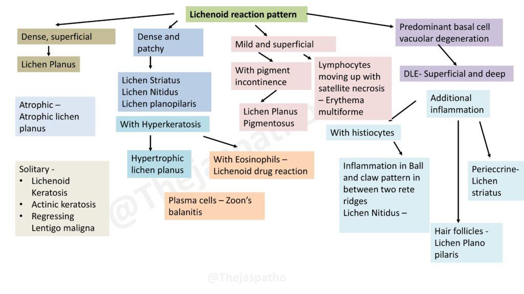
1. Lichenoid Reaction Pattern
The lichenoid reaction pattern is characterized by an inflammatory response that primarily targets the dermoepidermal junction. It features basal cell vacuolar degeneration with dense inflammatory infiltrates. This algorithm helps distinguish between various lichenoid conditions like Lichen Planus, Lichenoid Drug Reaction, and Lichen Striatus based on the presence of additional features such as eosinophils, histiocytes, or involvement of specific structures like hair follicles.
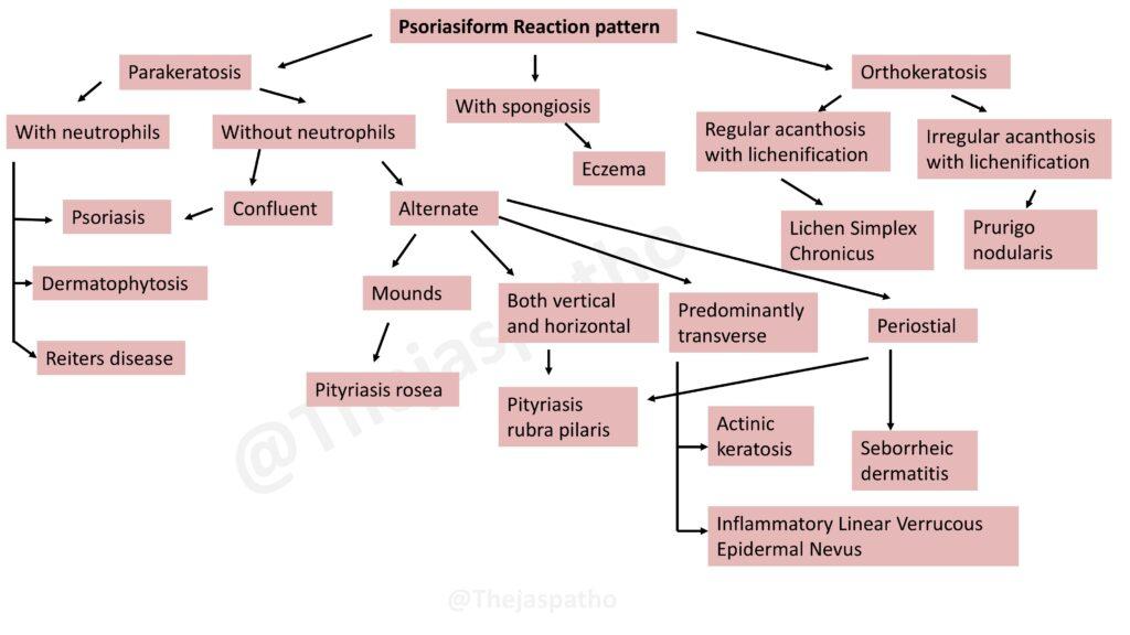
 2. Psoriasiform Reaction Pattern
This pattern is defined by epidermal hyperplasia, often with parakeratosis and acanthosis. The algorithm aids in differentiating between conditions such as Psoriasis, Seborrheic Dermatitis, and Lichen Simplex Chronicus, focusing on the arrangement of neutrophils, the pattern of parakeratosis, and the presence of spongiosis. It also addresses how to identify the involvement of fungal infections like Dermatophytosis.
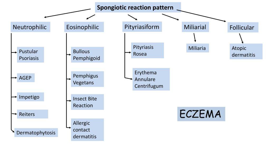
3. Spongiotic Reaction Pattern
Spongiosis, characterized by intercellular edema in the epidermis, is the hallmark of this reaction pattern. The algorithm guides you through distinguishing various spongiotic dermatoses such as Eczema, Allergic Contact Dermatitis, and Pityriasis Rosea. It also includes specific conditions that present with neutrophilic or eosinophilic infiltrates.
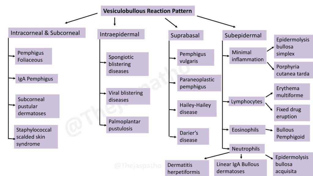
4. Vesiculobullous Reaction Pattern
The vesiculobullous pattern is identified by the formation of blisters at different levels of the epidermis. This algorithm categorizes diseases like Pemphigus Foliaceous, Bullous Pemphigoid, and Dermatitis Herpetiformis based on the blister location (intracorneal, subcorneal, intraepidermal, suprabasal, or subepidermal) and the type of inflammatory cells present.
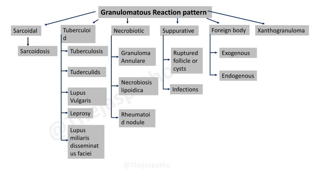
5. Granulomatous Reaction Pattern
Granulomatous inflammation involves the presence of granulomas within the dermis. This algorithm offers a structured approach to identifying different types of granulomatous diseases, such as Sarcoidosis, Leprosy, and Granuloma Annulare, based on their morphology (sarcoidal, tuberculoid, necrobiotic, or suppurative) and associated features.
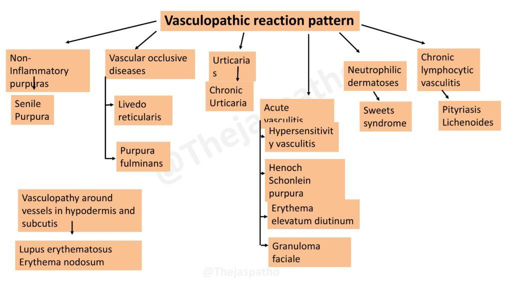
6. Vasculopathic Reaction Pattern
This pattern represents a range of vascular conditions, from non-inflammatory purpuras to vasculitis and neutrophilic dermatoses. The algorithm differentiates between entities like Henoch-Schönlein Purpura, Hypersensitivity Vasculitis, and Lupus Erythematosus by examining the inflammatory infiltrate, vessel involvement, and localization of the vascular changes.

