

Giant Cell Arteritis (Temporal Arteritis): Pathogenesis, Pathology and Clinical Features
Giant cell arteritis, also known as temporal arteritis, is a form of large-vessel vasculitis that primarily affects elderly individuals. It commonly involves large and medium-sized arteries, especially the branches of the external carotid artery, with the temporal artery being the most frequently affected.
Early recognition and prompt treatment of this condition are essential as it has high risk of vision loss.
Epidemiology
Giant cell arteritis typically affects individuals above the age of 50 years. It is one of the most clinically significant vasculitides seen in elderly patients.
The disease primarily involves large and medium-sized arteries.
The most commonly affected vessels include:
Temporal artery
Branches of the external carotid artery
Ophthalmic artery (involvement is particularly important because it may lead to sudden visual loss).
Pathogenesis
Giant cell arteritis is characterized by granulomatous inflammation of the arterial wall.
The inflammatory process involves:
Activation of immune cells within the vessel wall
Infiltration of inflammatory cells and
Formation of multinucleated giant cells
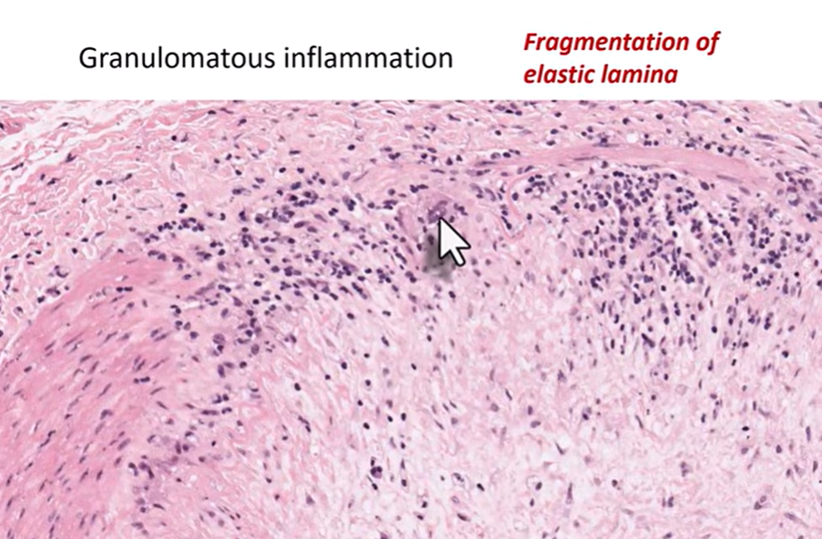
The hallmark microscopic finding is granulomatous inflammation of the arterial wall
Characteristic features include:
Presence of multinucleated giant cells
Fragmentation of the internal elastic lamina
Inflammatory infiltrate within the vessel wall
Intimal thickening
Luminal Narrowing
This luminal narrowing can significantly reduce blood flow, leading to ischemic complications.
Clinical Features :
Patients with giant cell arteritis may present with a variety of symptoms, due to reduced blood supply to tissues supplied by affected arteries. These include
Headache
Scalp tenderness
Jaw claudication
Visual disturbances
Major Complication: One of the most serious complications of giant cell arteritis is sudden loss of vision.
This occurs when the ophthalmic artery becomes involved, leading to reduced blood supply to the optic structures.
Watch the Full Lecture
Students can watch the complete explanation of this topic in the lecture below.
