

Gross Pathology Specimen Series – Part 3
A continuation of our visual learning series featuring classic specimens encountered in pathology. Each image is labeled and described to aid easy revision and practical understanding.
All images are optimized for fast viewing and learning, whether you’re revising on your laptop or mobile.
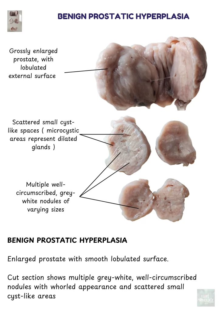
Benign Prostatic Hyperplasia
Gross Description:
Enlarged prostate with a smooth lobulated surface. Cut surface shows multiple well-circumscribed grey-white nodules and scattered cyst-like areas representing dilated glands.
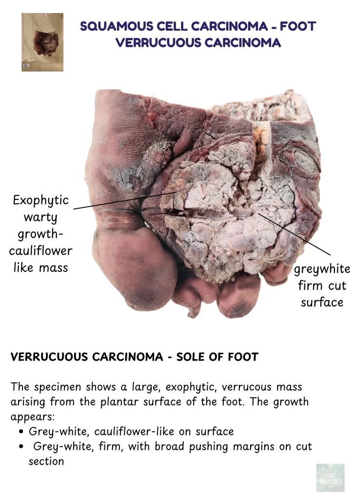
Verrucous Carcinoma – Sole of Foot
Gross Description:
A large exophytic warty mass arising from the plantar aspect of the foot. The lesion is grey-white, cauliflower-like on surface
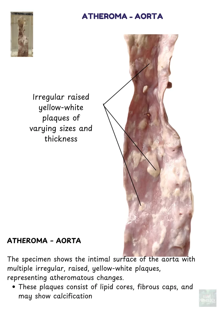
Atheroma – Aorta
Gross Description:
Multiple raised, yellow-white plaques of varying size and thickness on the intimal surface of the aorta. These represent atherosclerotic changes, with lipid cores, fibrous caps, and possible calcification.
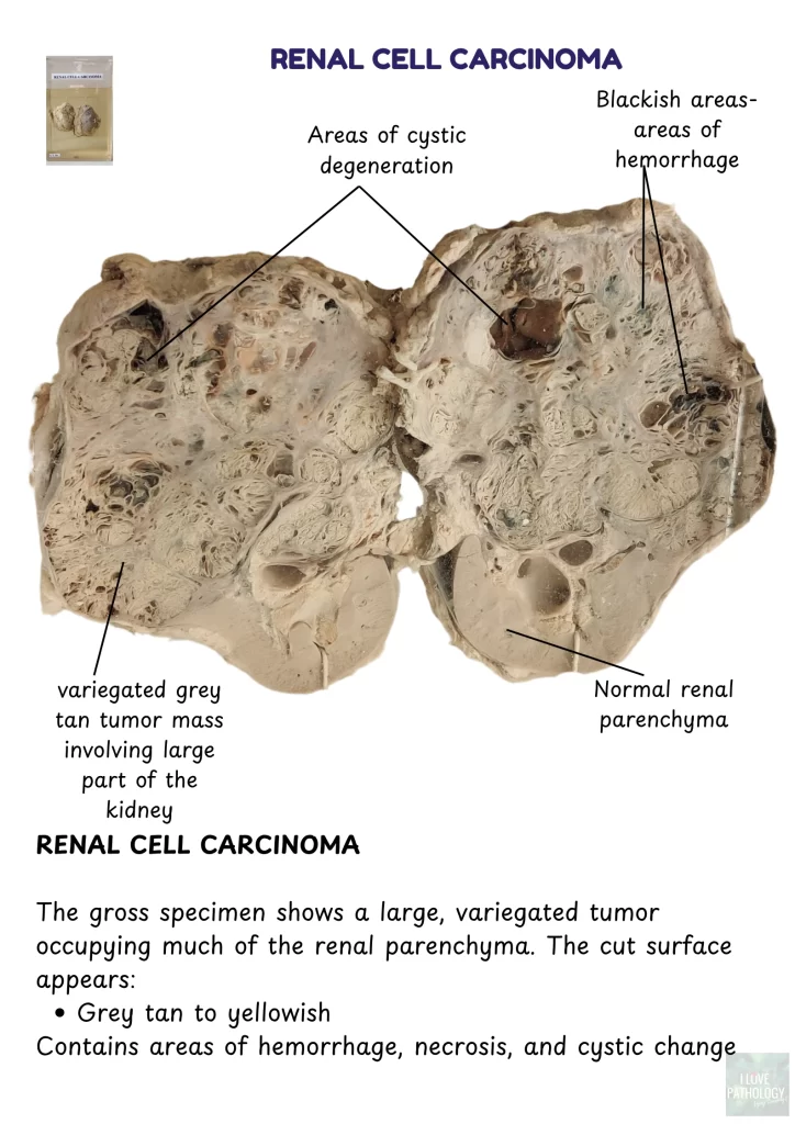
Renal Cell Carcinoma
Gross Description:
Large variegated grey-tan tumor replacing much of the renal parenchyma. Shows areas of cystic change, hemorrhage, and necrosis.
🧬 Leiomyoma – Uterus
Gross Description:
Well-circumscribed firm white mass with a whorled appearance, arising from the myometrium. Can be submucosal, intramural, or subserosal depending on location.

“This post is part of an ongoing series to help students identify gross pathology specimens quickly for theory and practical exams.”