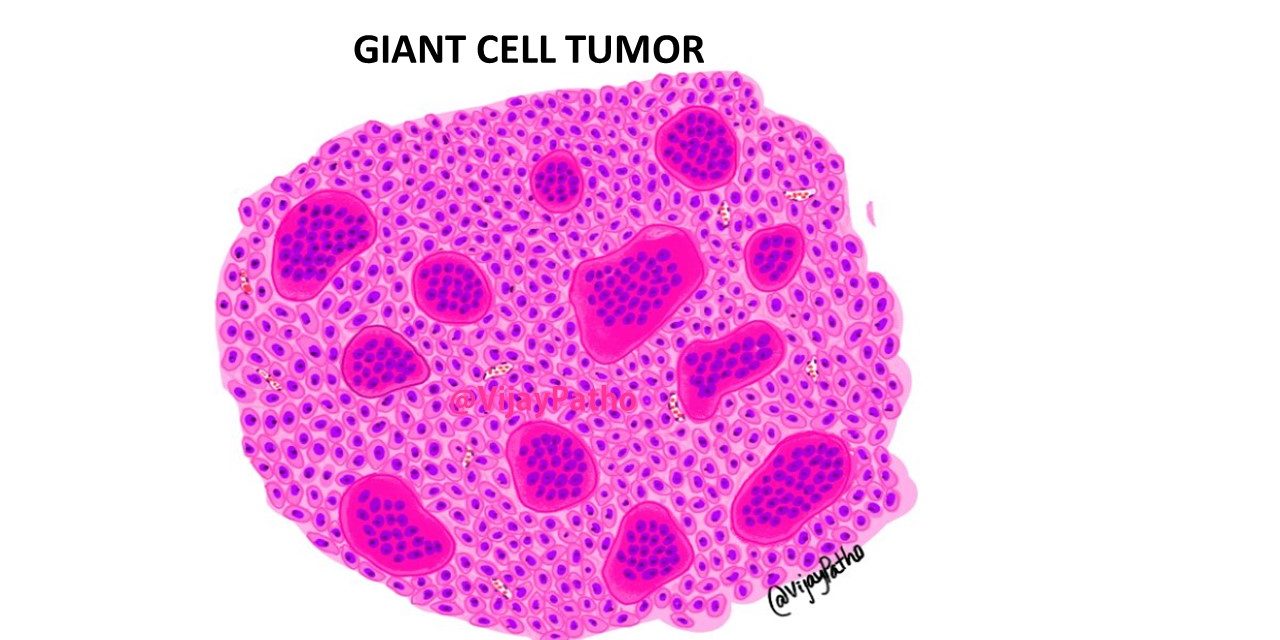
Giant cell tumor (GCT) also referred to as osteoclastoma is a locally aggressive neoplasm characterized by large numbers of osteoclast-type giant cells, uniformly distributed in a population of mononuclear plump epitheloid or spindle cells.
GCTs are more common in 2nd to 3rd decades of life and these are epiphyseal, eccentric and expansile lesions.
Most common location is around knee joint i.e proximal part of tibia and distal part of femur. Distal radius, sacrum can also be involved.
Radiologically these are lytic lesions, eccentrically placed, epiphyseal in location,which expands and destroys the overlying cortex. Lesions extending into the soft tissue often have a thin rim of bone ( “egg-shell” appearance) or if multiloculated have “soap-bubble” appearance.
Gross findings: can markedly vary
May be red, hemorrhagic and fleshy tumor, usually large and appear variegated. Extensive cystic degeneration can also be seen.
Microscopic findings:
Giant cell tumor consists of two different populations
1. Mononuclear cells: these are round to oval, occasionally spindled and contain round to oval nuclei with one to two prominent nucleoli.
2. Multinucleated giant cells: The giant cells have abundant eosinophilic cytoplasm and may contain numerous nuclei and these nuclei are identical to that to mononuclear cells.
The stroma can contain thin walled capillaries, areas of hemorrhage can rarely be seen along with hemosiderin laden macrophages. Areas of cystic degeneration , mitotic figures and rarely necrosis can be seen.


