
It is a chronic granulomatous inflammation of the lymph node with the presence of caseation necrosis. It is caused by infection with Mycobacterium tuberculosis.
It is one of the most common extrapulmonary manifestations of tuberculosis.
TB lymphadenitis may occur due to either of the following reasons
1. Spread from the infections of tonsil to cervical lymph nodes
2.Reactivation of healed focus which was involved during primary infection
3.Spread from the lung to the mediastinal lymph nodes
4.Hematogenous spread as in the case of military tuberculosis
The most common presentation is nontender enlargement of lymphnodes present for long duration. In cervical group of lymphnode enlargement, porterior triangle group of lymph nodes are most commonly affected, followed by anterior triangle, submandibular and supraclavicular group of lymph nodes.
If multiple lymphnodes are involved and as they enlarge, they adhere to one another because of periadenitis (inflammation of the tissue surrounding the lymphnode) and are referred to as “Matted” group of lymph nodes.
Complications include ulceration, fistula, or abscess formation. An abscess which occurs as a complication of tuberculosis is referred to as “cold abscess”, as there is no or little signs of inflammation.
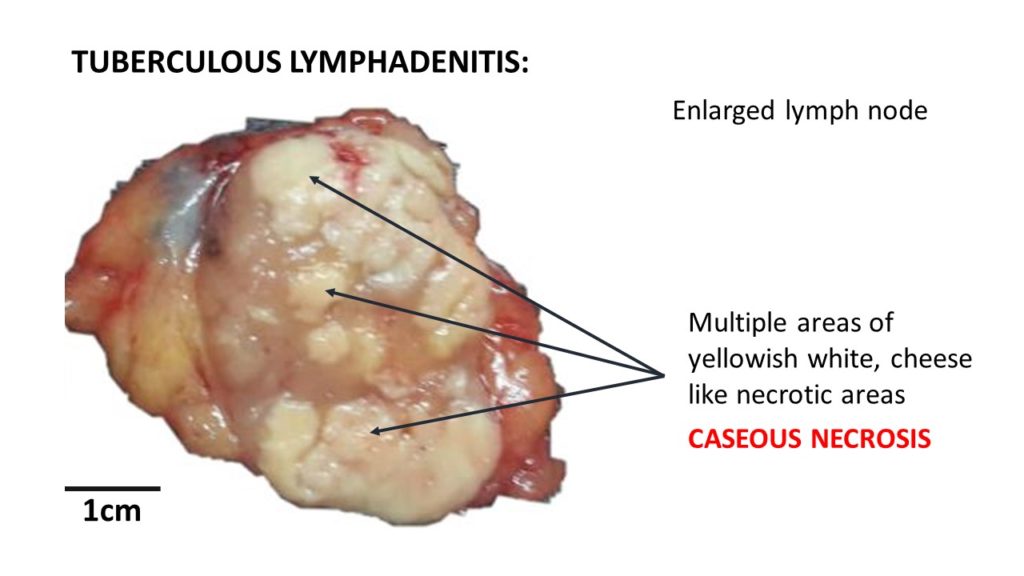
Gross: the lymph nodes will be enlarged and matted. Cut section reveals single or multiple areas of cheesy yellowish material which is called as caseous necrosis ( Cheese- like).
Microscopy:
The characteristic feature is presence of granulomas with presence of caseous necrosis.
The granuloma is collection of modified macrophages which resemble “epithelial” cells and hence are referred to s “epithelioid cells”. These cells have abundant pale cytoplasm with pale oval, elongated or slipper shaped nuclei.
Some of these epitheliod cells fuse together to form multinucleated giant cells which are refereed to as Langhans type of Giant cells.
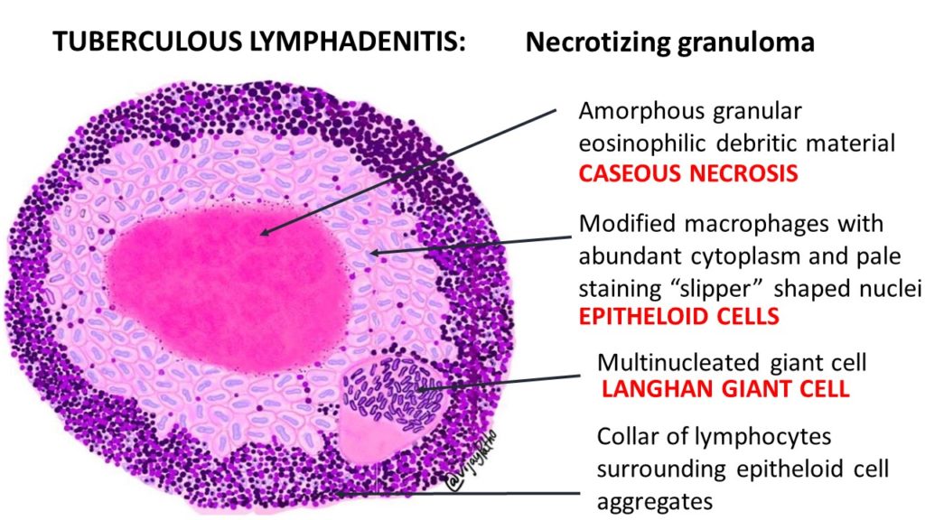
Caseous necrosis is evident in the centre of these granulomas. Caseous necrosis on H & E staining appears as amorphous granular eosinophilic debritic material.
Collar of lymphocytes are seen surrounding these epitheloid cell aggregates. In the older lesions evidence of fibrosis is also seen surrounding these granulomas, beyond the lymphoid cuff.