Congestion Versus Hyperemia
Congestion: it is a passive process which results due to impaired outflow of venous blood from a tissue
Should not be confused with hyperemia. Hyperemia is an active process which results due to dilatation of arterioles and increased inflow of blood as in sites of inflammation.
CHRONIC VENOUS CONGESTION – LUNG
‘The most common mechanism for CVC lung is left sided heart failure. The Left heart failure many be due to coronary heart disease or long standing hypertension”
Gross Morphology: The lungs are heavy and is rusty brown colored on cut section as a result of hemosiderin laden macrophages. Due to fibrosis, the lungs are firm in consistency. The combination of brown color and firmness resulted in the name “ brown induration” of lung.
Microscopy:
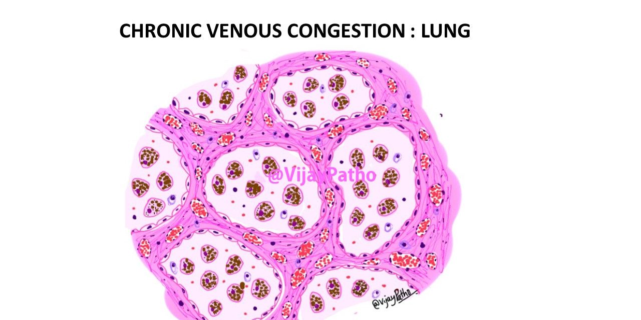
The alveolar wall show dilated and congested capillaries and it is markedly thickened due to increase in the fibrous connective tissue.
The alveolar spaces contain numerous hemosiderin laden macrophages which are also referred to as “heart failure” cells. These heart failure cells can also be seen in the septa occasionally.
Heart failure cells: These are basically hemosiderin laden macrophages/siderophages.
Due to passive congestion with dilated capillaries, the rbc’s leak into the alveolar spaces and are broken down resulting in release of hemoglobin. This hemoglobin is phagocytosed by the alveolar macrophages where it is degraded to release hemosiderin and biliverdin. The hemosiderin accumulates in the form of golden brown pigment as more and more rbc’s are lysed. Since these pigment laden macrophages are seen in the setting of heart failure, these are sometimes called as “ HEART FAILURE” cells.


