Testicular tumors are rare, accounting for only 1% of all neoplasms. However, they are the most common type of neoplasm in young adults. This blog will guide you through the anatomy, histology, and the latest WHO classification of testicular tumors.
What are the basic anatomical features of the testes?
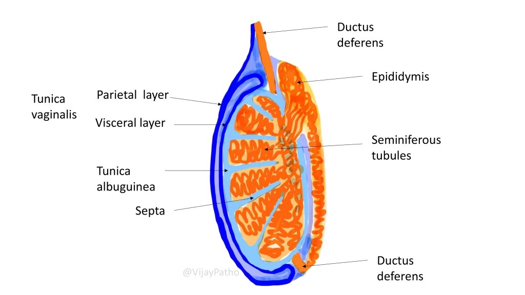
The testes are paired, oval-shaped organs located in the scrotum. They measure approximately 4-5 cm in length, 2.5 cm in width, and about 3 cm in thickness. The average weight of each testis is around 15-20 grams.
The testes contain seminiferous tubules, where sperm production occurs. They are surrounded by connective tissues called septa, which divide the testicular parenchyma into compartments. The outer covering of the testes includes:
Tunica albuginea: The thick connective tissue covering.
Tunica vaginalis: A double-layered membrane with parietal and visceral layers.
What are the histological components of the testes?
The histology of the testes shows:
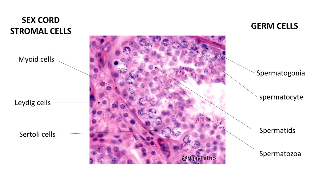
Seminiferous tubules: Lined by germ cells and Sertoli cells (support cells).
Interstitial cells: Predominantly Leydig cells, responsible for testosterone production.
How are the cells of the testes classified histologically?
Germ cells: Found within the seminiferous tubules, undergoing various stages of maturation from spermatogonia to mature spermatozoa.
Sex cord-stromal cells: Includes Sertoli cells (supporting the germ cells) and Leydig cells (producing testosterone).
Functions of Testes
What are the primary functions of the testes?
The testes have two major functions:
Reproductive function: Involves spermatogenesis, the production of male gametes (spermatozoa), occurring within the seminiferous tubules.
Endocrine function: Includes the production of testosterone by Leydig cells. Testosterone regulates secondary sexual characteristics, libido, and spermatogenesis. Sertoli cells also secrete inhibin, which provides negative feedback to the anterior pituitary, suppressing follicle-stimulating hormone (FSH) secretion.
Testicular Tumors
How common are testicular tumors?
Testicular tumors account for:
1% of all adult neoplasms.
5% of urological tumors.
They are most common in young men aged 15-40 years. Most testicular tumors have an indolent (slow-growing) course.
How are testicular tumors classified?
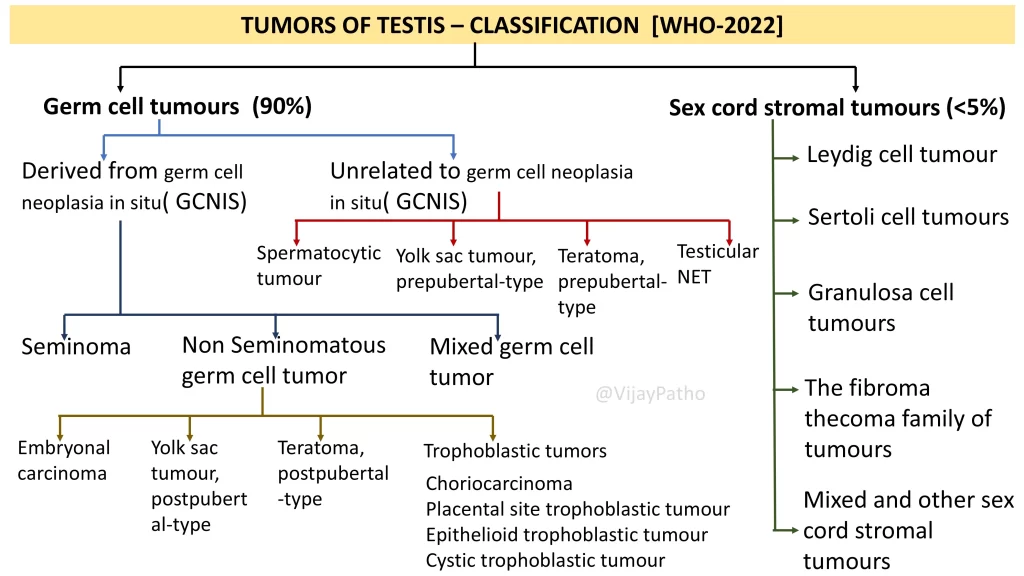
According to the latest WHO classification, testicular tumors are divided into:
Germ cell tumors: Constitute around 90% of all testicular tumors.
Sex cord-stromal tumors: Constitute less than 5% of testicular tumors.
What are the subtypes of germ cell tumors?
Germ cell tumors are further categorized into:
Derived from germ cell neoplasia in situ (GCNIS): Includes seminomas, non-seminomatous germ cell tumors, and mixed germ cell tumors.
Non-seminomatous germ cell tumors: Include embryonal carcinoma, yolk sac tumor (post-pubertal type), teratoma (post-pubertal type), and trophoblastic tumors (e.g., choriocarcinoma, placental site trophoblastic tumor).
Unrelated to GCNIS: Includes spermatocytic tumors, yolk sac tumor (pre-pubertal type), and testicular neuroendocrine tumors.
What are the subtypes of sex cord-stromal tumors?
Sex cord-stromal tumors are classified into:
Leydig cell tumors: Derived from the interstitial Leydig cells, responsible for testosterone production.
Sertoli cell tumors: Originating from Sertoli cells, which support germ cells.
Granulosa cell tumors and fibroma-thecoma family tumors: Rare tumors that share a common embryological origin with ovarian tumors due to the shared gonadal ridge ancestry.
WHO Classification of Testicular Tumors
Why do granulosa cell tumors occur in testes?
The testes and ovaries share a common embryological origin from the gonadal ridge. While the testes differentiate into sex cords and stromal cells, some progenitor cells remain pluripotent, leading to granulosa or fibroma-like tumors in the testes.
click below to watch the video on classification of testicular tumors

