SYSTEMIC LUPUS ERYTHEMATOSUS (SLE)
SLE is the prototype of a multisystem disease of autoimmune origin, characterized by a vast array of autoantibodies, particularly antinuclear antibodies (ANAs) in which injury is caused mainly by deposition of immune complexes and binding of antibodies to various cells and tissues
Clinically: Chronic, and relapsing, often febrile illness characterized principally by injury to the skin, joints, kidney, and serosal membranes
Etiology:
Fundamental defect in SLE is a failure of the mechanisms that maintain self-tolerance.
• Genetic Factors –
1. Family members of patients have an increased risk of developing SLE (20% of clinically unaffected first-degree relatives of SLE patients reveal autoantibodies and other immunoregulatory abnormalities)
2. Higher rate of concordance (>20%) in monozygotic twins.
3. HLA associations – HLA-DQ locus have been linked to the production of anti–doublestranded DNA, anti-Sm, and antiphospholipid antibodies
4. Inherited deficiencies of early complement components, such as C1q , C2 or C4
5. Polymorphism in the inhibitory Fc receptor leading to inadequate control of B cell activation
• Environmental Factors – UV irradiation, Cigarette smoking, Sex hormones, Drugs (procainamide and hydralazine)
• Immunologic abnormalities –
1. Type 1 Interferons – for unknown reasons abnormally large amounts IFN alpha are produced
2. TLR signals – that recognize RNA and DNA, activate B cells specific for nuclear antigens
3. Failure of B cell tolerance – high frequency of auto-reactive B cell production compared to normal individuals
4. CD4 helper T cells, which escape tolerance – production of high affinity auto-antibodies
Pathogenesis:
The pathogenesis of systemic lupus erythematosis involves various steps which is simplified in the illustration below
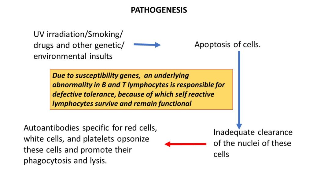
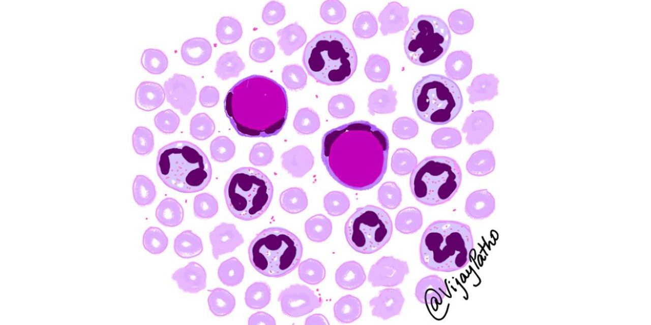
MECHANISM OF TISSUE INJURY: In tissues, nuclei of damaged cells react with ANAs, lose their chromatin pattern, and become homogeneous, to produce so-called LE bodies or hematoxylin bodies
CLINICAL FEATURES:
• Typically, the patient is a young woman with the following features:
a butterfly rash over the face, fever, pain but no deformity in one or more peripheral joints (feet, ankles, knees, hips, fingers, wrists, elbows, shoulders), pleuritic chest pain, and photosensitivity.
Other features:
DISCOID RASH: Erythematous raised patches with adherent keratotic scaling and follicular plugging; atrophic scarring may occur in older lesions.
PHOTOSENSITIVITY: Rash as a result of unusual reaction to sunlight.
ORAL ULCERS: Oral or nasopharyngeal ulceration, usually painless
ARTHRITIS: Nonerosive arthritis involving two or more peripheral joints, characterized by tenderness, swelling, or effusion.
SEROSITIS: Pleuritis and pericarditis.
RENAL MANIFESTATIONS: range from mild hematuria and proteinuria to acute renal insufficiency and may become the predominant cause leading to mortality
NEUROLOGIC INVOLVEMENT: Seizures and Psychosis due to antibodies against synaptic membrane protein. Acute vasculitis of small vessels noted.
To Read lab diagnosis and morphology of various organs in SLE





Recent Comments