
Hi All
I am overwhelmed by the response i received for the first five parts of “Similes & Metaphors of Pathology”. Thanks for the suggestions and giving me new clues/ similes. This has been an amazing learning experience for me too. I have decided to continue learning! I am planning to continue this series and will try to keep you engaged.
The Part 6 of ” Similes & Metaphors of Pathology” is here. ENJOY LEARNING 🙂
Note: Mobile users please use landscape mode for complete view.
More interesting ones to come in Part 7!….

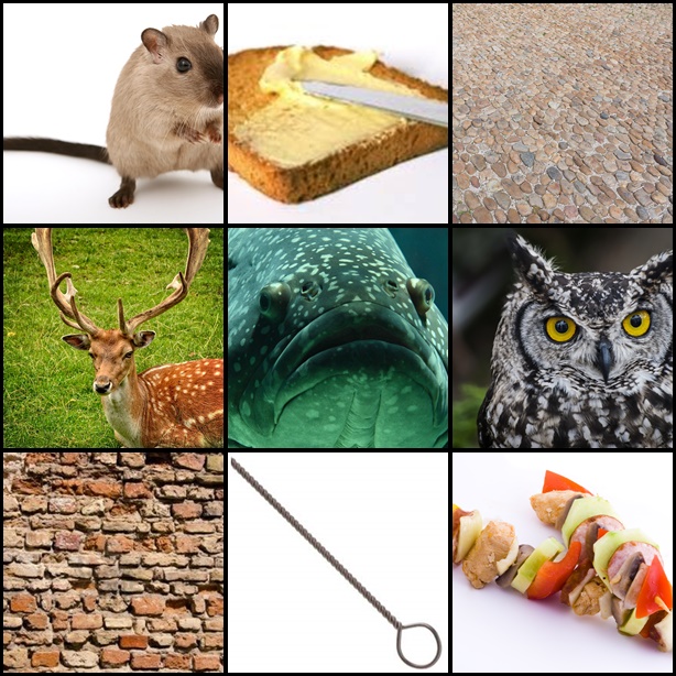
Rodent |
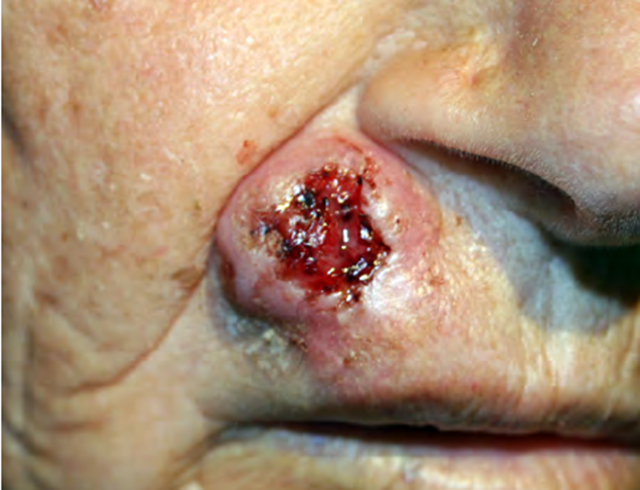
Rodent Ulcer- Basal cell Carcinoma ( source) |
Basal cell carcinoma- Rodent ulcer.
Rodent: mammals with pairs of continuously growing, razor-sharp incisors. from Latin rodere, “to gnaw” ulcer has an indurated edge and ulcerated centre. It is slow growing but spreads deep This is commonly known as rodent ulcer. Because of its “Gnawed” appearance i.e.to bite or chew/make a hole by chewing! |

Bread and Butter |

Bread Butter appearance- Fibrinous pericarditis ( source ) |
Fibrinous pericarditis- bread and butter pericarditis.
It’s an exudative inflammation. In acute stage of Rheumatic heart disease, there will be tenacious deposits of fibrin in the pericardium which resembles that of irregular surface of the buttered bread/ Bread butter appearance. Also seen in uremic pericarditis |

Cobble stone |
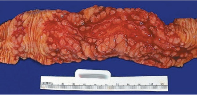
Crohns disease- cobble stone appearance.(S0urce) |
Crohns disease- cobble stone appearance.
It’s a morphological appearance Islands of intact, edematous mucosa which is surrounded by ulcers(Communicating fissures and crevices in the mucosa)s how the classic “cobble stone appearance. |

Antler |

Fibroadenoma: Antler horn configuration Source |
Antlers are a pair of bony, branched structures that protrude from the skull of animals
Fibroadenoma: Antler horn configuration of ductal epithelial cells. This is the finding seen in smears prepared from fine needle aspiration of fibroadnoma. The aspirates will be hypercellular with monolayerd sheets of ductal epithelial cells. These sheets are described as staghorn or antler horn configuration on the edges of these cellular sheets. |

Diladipated Bricks |

Hailey Hailey disease: Diladipated Brick appearance. Source |
Hailey Hailey disease: Diladipated Brick appearance.
This disease is an autosomal dominant disorder with recurrent eruption of vesicles and bullae. There will be suprabasal separation resulting in blister. The intercellular bridges are lost leading to acantholysis, with few intact intercellular bridges results in “Diladipated Brick appearance”
|
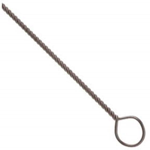
Wire Loop |
 Lupus nephritis- wire loop lesions ( Source) Lupus nephritis- wire loop lesions ( Source) |
Lupus nephritis- wire loop lesions
It is a light microscopic finding in renal biopsy in Lupus nephritis where the capillary walls are thickened due to subendothelial immune complex deposits. capillaries stains deep red and appears as homogenous and “rigid” thickening of peripheral capillary loops. This change is called the “wire loop” lesion. |

Shish-Kebab |

Candidiasis- Pap smear finding- Shish-Kebab appearance (source) |
Candidiasis- Pap smear finding- Shish-Kebab appearance
The candida are long filamentous organisms. The squamous epithelial cells, peculiarly aggregate around these filamentaous organisms, appears as if these cells are speared by these pesudohyphae, giving Shish- kebab appearance. |

Fish mouth |

Rheumatic heart disease: Fish mouth appearance ( Source) |
Rheumatic heart disease: Fish mouth appearance of Mitral valve.
Most of the patients with rheumatic valvular heart disease have an affected mitral valve. There will be severe mitral stenosis due to marked thickening and fibrosis.This results in narrowing of its orifice to a slit, which resembles Fish mouth. |
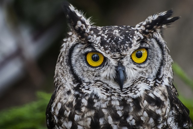
Owl Eye |
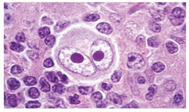
Reed Sternberg Cell ( Source ) |
Hodgkin lymphoma: Owl’s eye appearance of nucleus
The characteristic features of the nuclei of Reed Sternberg cells in Hodgkin disease. These cells have symmetric mirror image bilobed nucleus which resembles that of owl’s eye. The intranuclear inclusions in Cytomegalovirus-infected cells also looks like owl’s eye. |
How did you like this part of ” Similes & Metaphors in Pathology”? Have you come across any interesting similes/metaphors in teaching Pathology?
Feel free to comment and give your suggestions below or mail me at vijayshankar@ilovepathology.com

