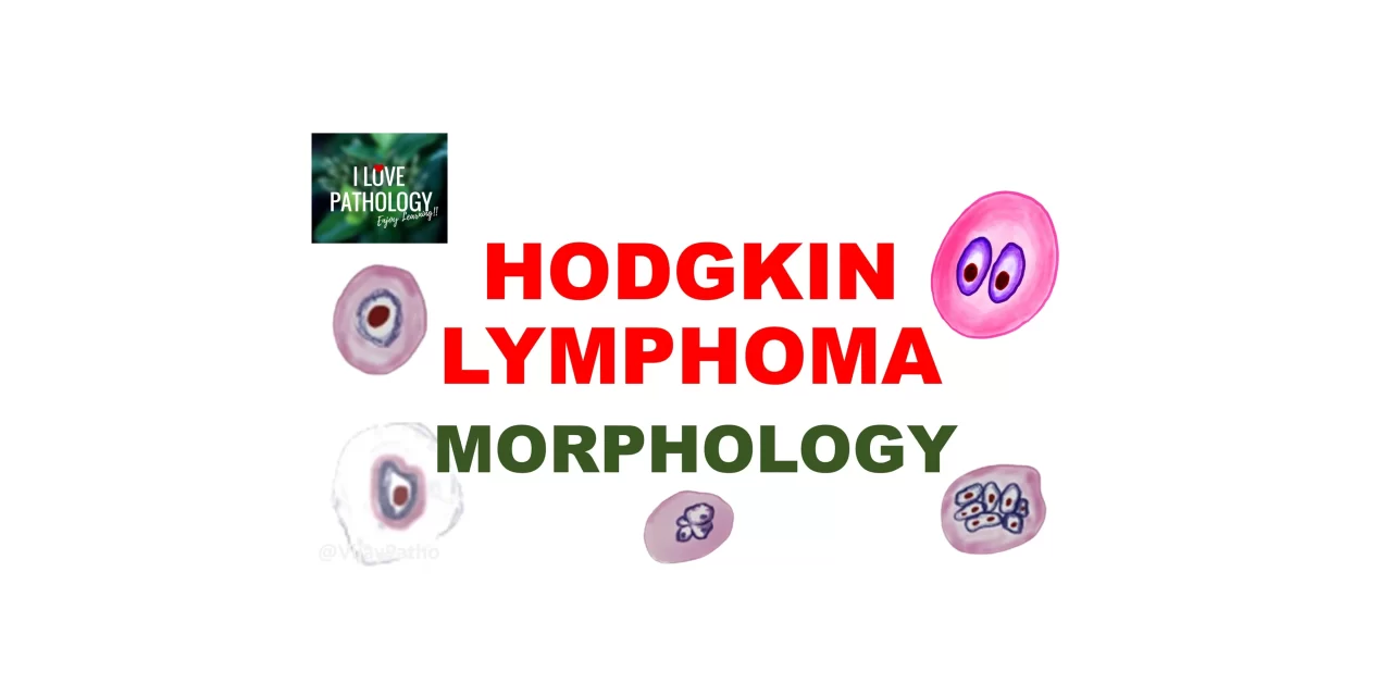
Hodgkin Lymphoma: Morphology and Reed-Sternberg Cell Variants
What are the two major types of Hodgkin lymphoma?
Hodgkin lymphoma is classified into:
Classical Hodgkin Lymphoma (CHL)
Nodular Lymphocyte Predominant Hodgkin Lymphoma (NLPHL)
What are the subtypes of classical Hodgkin lymphoma?
Classical Hodgkin lymphoma includes:
Nodular sclerosis
Mixed cellularity
Lymphocyte-rich
Lymphocyte-depleted
What is essential to diagnose Hodgkin lymphoma?
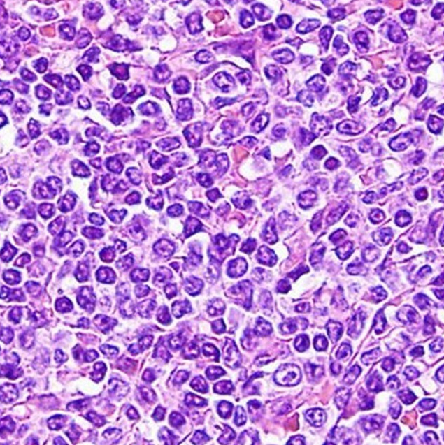
The diagnosis requires identifying Reed-Sternberg (RS) cells or their variants, especially within a background of non-neoplastic inflammatory cells.
What are the variants of Reed-Sternberg cells?
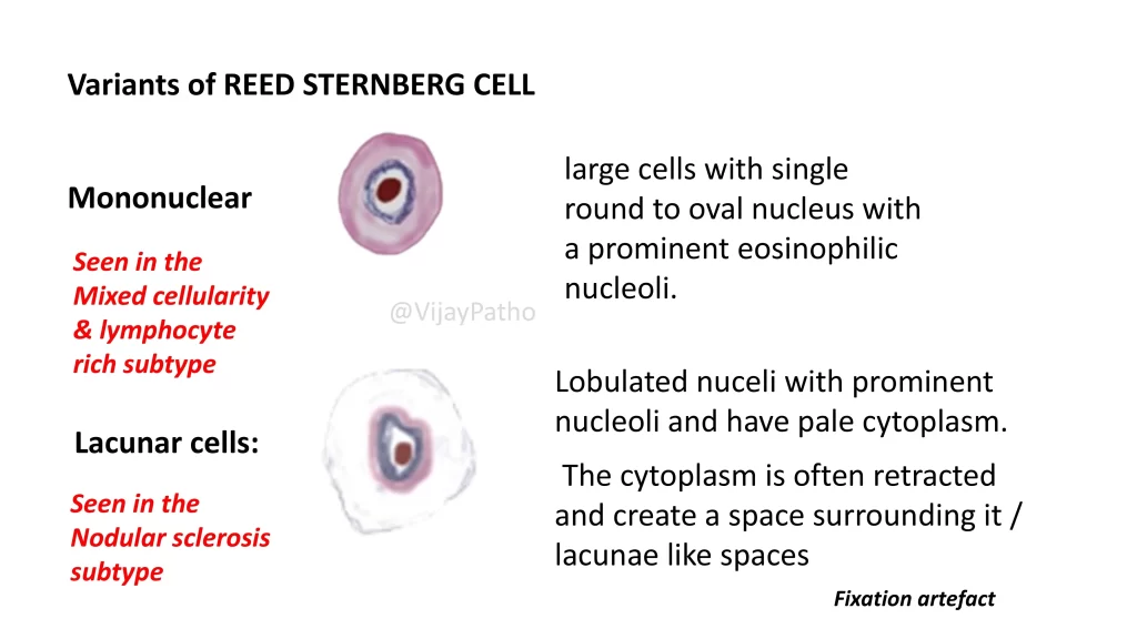
Mononuclear variant:
A large cell with a single round or oval nucleus and a prominent eosinophilic nucleolus. Found in mixed cellularity and lymphocyte-rich types.
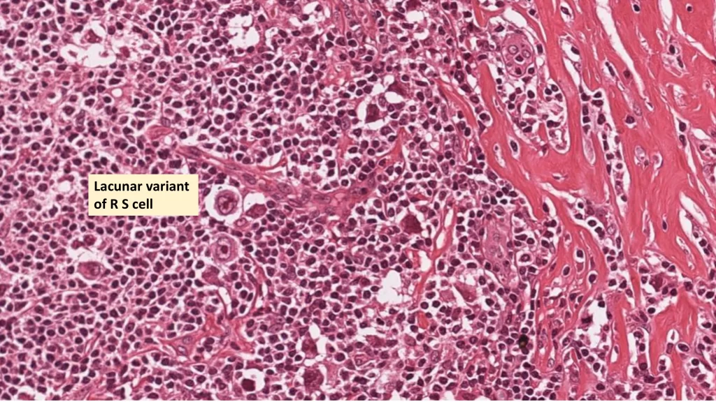
Lacunar variant:
Characterized by a lobated nucleus and retracted pale cytoplasm, forming a clear space (lacuna) around it. This is a fixation artifact and is common in the nodular sclerosis subtype.
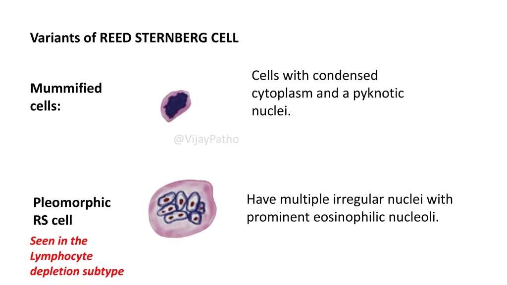
Mummified cell:
These cells show condensed cytoplasm and pyknotic nuclei.
Pleomorphic RS cells:
These cells have multiple irregular nuclei with prominent eosinophilic nucleoli and are seen in lymphocyte-depleted subtype.
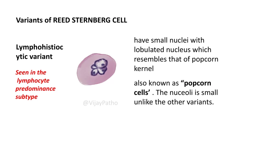
Lymphohistiocytic variant (Popcorn cell):
Features a lobulated small nucleus, resembling a popcorn kernel. It lacks prominent nucleoli and is found in nodular lymphocyte predominant Hodgkin lymphoma.
RS-like cells can appear in conditions like infectious mononucleosis, some solid tumors, and large cell non-Hodgkin lymphomas. Immunophenotyping helps differentiate true RS cells from look-alikes.
Subtypes of Classical Hodgkin Lymphoma
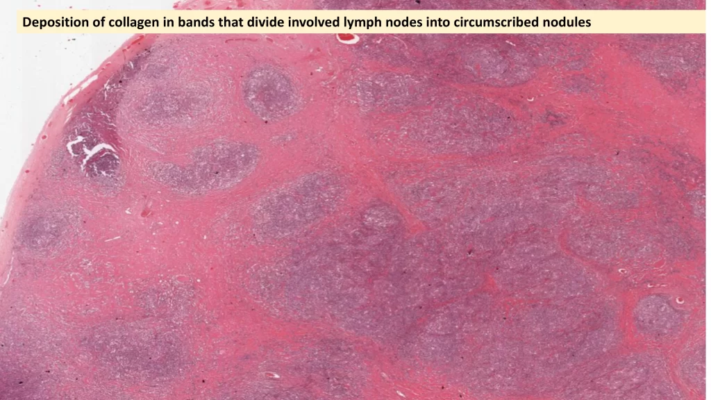
Nodular sclerosis : is the most common, accounting for 65–70% of cases.
Features of nodular sclerosis?
Presence of lacunar RS cells
Collagen bands divide the lymph node into nodules
Rarely associated with Epstein-Barr virus (EBV)
Immunophenotype: Positive for Pax5, CD15, and CD30; Negative for B-cell and T-cell markers, and CD45 (leukocyte common antigen)
Mixed cellularity subtype
Accounts for 20–25% of cases
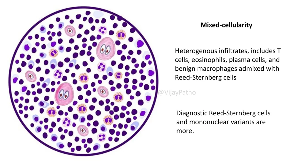
Diffuse infiltration of the lymph node by a mixed inflammatory background including eosinophils, plasma cells, macrophages
Contains both classical and mononuclear RS cells (mononuclear variant is also called Hodgkin cell)
EBV association in ~70% of cases
Lymphocyte-rich subtype
Majority of cells are reactive lymphocytes
Lymph node may appear nodular due to residual B-cell follicles
RS and mononuclear variants seen
Classic immunophenotype (Pax5+, CD15+, CD30+, negative for B/T cell markers)
EBV associated in ~40% of cases
Prognosis: Excellent
Lymphocyte-depleted subtype.
Rare
Shows very few lymphocytes and abundant pleomorphic RS cells
EBV association ~90%
More common in older adults, HIV-positive individuals, and in low-income countries
Prognosis is poor compared to other subtypes
Nodular Lymphocyte Predominant Hodgkin Lymphoma (NLPHL)
It’s an uncommon type, seen in about 5% of Hodgkin lymphoma cases.
Nodular architecture without collagen bands
Nodules contain small lymphocytes, histiocytes, and popcorn (lymphohistiocytic) cells
EBV is rarely associated
Retains B-cell markers like CD20 and BCL6
Negative for CD15 and CD30
IgH gene shows ongoing somatic hypermutation, indicating origin from transformed germinal center B cells
Quick Recap
Essential diagnostic cell: Reed-Sternberg cell
Lacunar cells: Nodular sclerosis
Most common subtype: Nodular sclerosis
Least common subtype: Lymphocyte-depleted
EBV association: Seen across types, most in lymphocyte-depleted
Classical HL immunophenotype: Pax5+, CD15+, CD30+, negative for CD20, CD45
NLPHL immunophenotype: CD20+, BCL6+, negative for CD15 and CD30
Popcorn cells: Seen in NLPHL
Click here to watch the video tutorial on morphology and R S variants