TRENDING:

Blood bags , types and uses
CHRONIC VENOUS CONGESTION – LUNG
Urine Analysis: Common Questions
Pathology Made Simple
  • Home
  • Education Contents
    • General Pathology
    • Systemic Pathology
    • Hematology
    • Clinical Pathology
    • Practical Pathology
      • Gross Pathology
  • Videos
  • Research
  • Pearls
  • Authors
  • About
    • Contact Me
    • Privacy Policy & Copyright notice
  • POSTGRADUATE
    • AUTOPSY

Select Page

Gross Pathology Specimen Series – Part 1

Posted by Dr Vijay Shankar S | Jun 27, 2025 | Practical Pathology, Gross Pathology |

Gross Pathology Specimen Series – Part 1

Welcome to Part 1 of the Gross Pathology Specimen Series — a visual learning resource for medical students and pathology enthusiasts. This series showcases classic and commonly encountered gross specimens, each accompanied by a short description to help you recognize key features, correlate with pathology, and prepare for practical exams or viva.

All images are optimized for fast viewing and learning, whether you’re revising on your laptop or mobile.
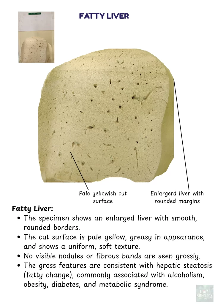
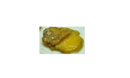

 Fatty Liver

Enlarged pale yellow liver with greasy texture and smooth borders.

fatty liver


 Chronic Venous Congestion – Liver (Nutmeg Liver)

Speckled cut surface resembling a nutmeg; red congested central veins and pale periphery.

CVC liver


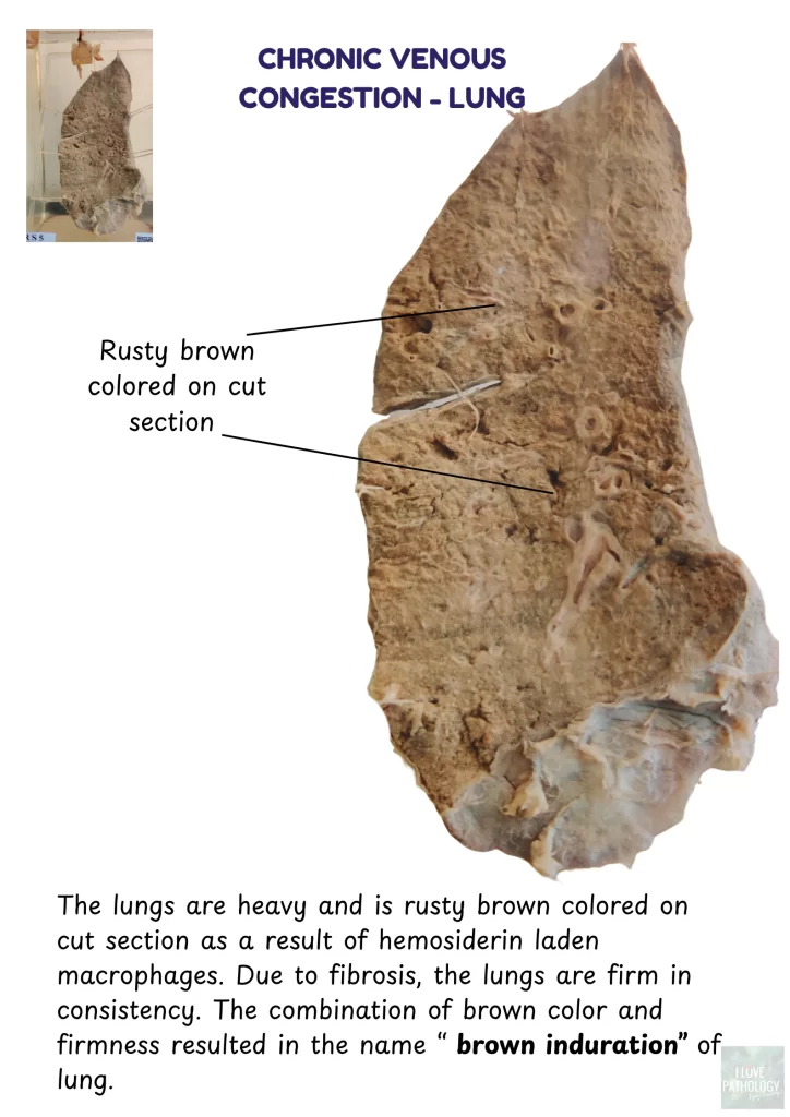
 Chronic Venous Congestion – Lung (Brown Induration)

Rusty brown cut surface due to hemosiderin-laden macrophages and fibrosis.

CVC lung


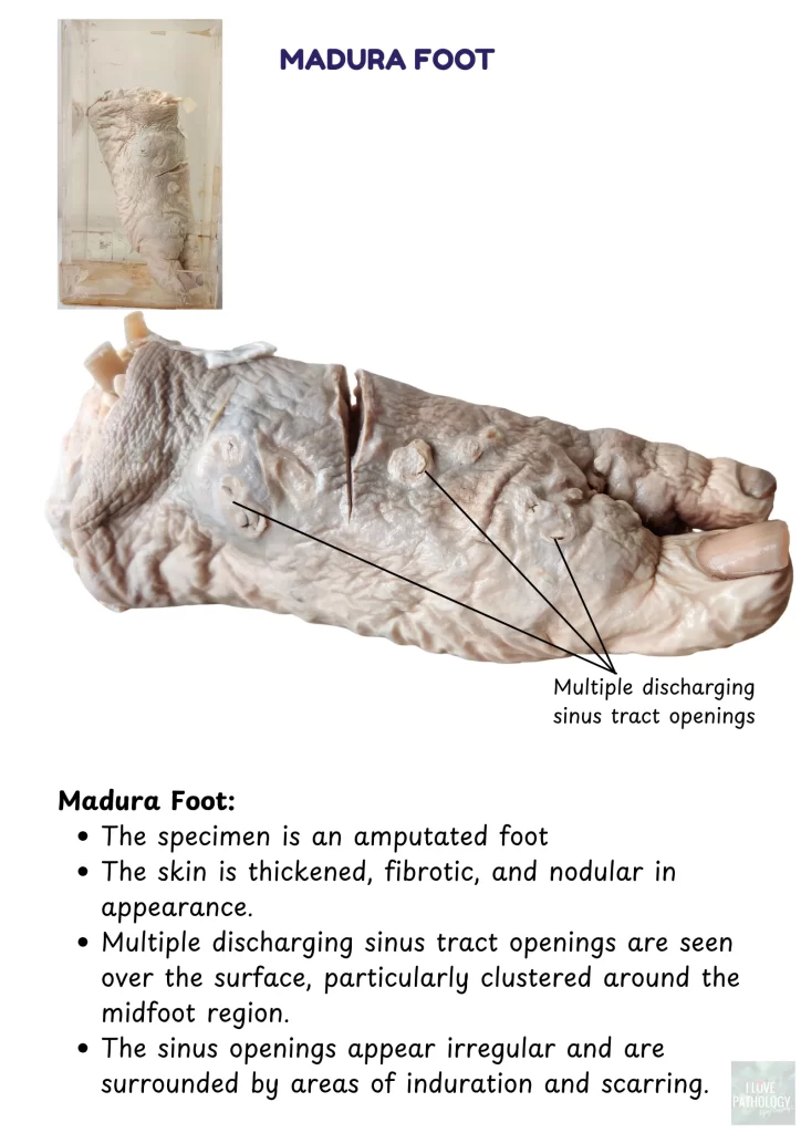
Madura Foot (Mycetoma)

Thickened fibrotic foot with discharging sinus tracts – typical of mycetoma.

madura foot


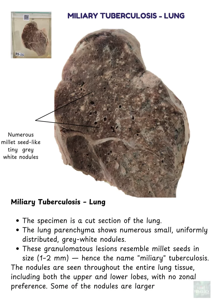
Miliary Tuberculosis – Lung

Numerous millet-sized grey-white nodules across the lung.

miliary tuberculosis


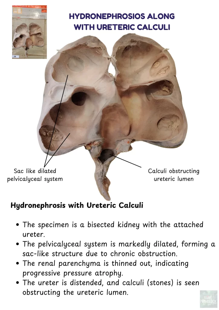
 Hydronephrosis with Ureteric Calculi

Sac-like dilated pelvicalyceal system and ureteric stones.

hydronephrosis


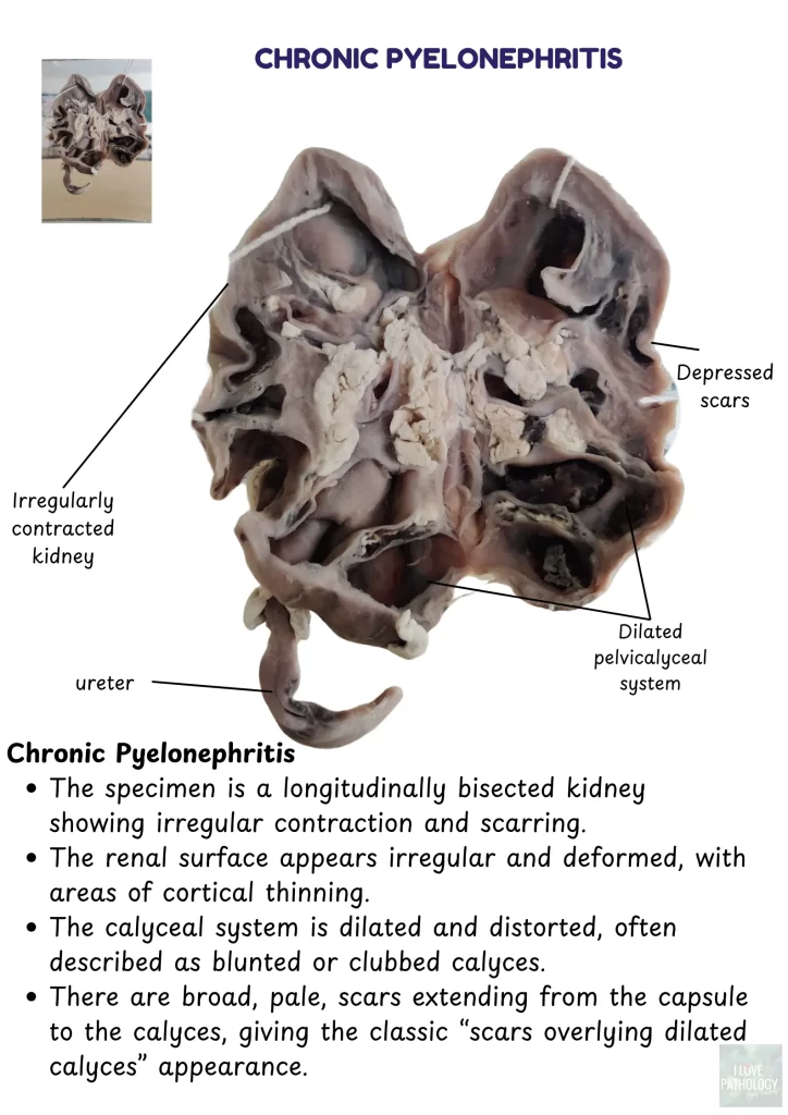
 Chronic Pyelonephritis

Scarred kidney with irregular surface and dilated calyces.

chronic pyelonephritis


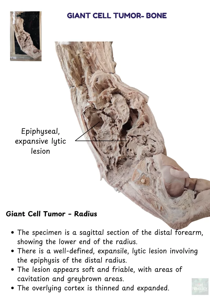
 Giant Cell Tumor – Radius (Bone)

Epiphyseal lytic lesion with cortical expansion and soft friable tissue.

Giant cell tumor - bone


Share:

Rate:

PreviousPneumonia: Etiopathogenesis and Classification 
NextWhat to Know Before Visiting a Cannabis Dispensary for the First Time

About The Author

Dr Vijay Shankar S

Dr Vijay Shankar S

With more than 20 years of experience as an academician, He has developed the art of content generation to make the learning more fun. Loves to promote social media for education in Pathology

Related Posts

Pathology of Schwannoma

Pathology of Schwannoma

June 27, 2020

Pathology of Lipoma

Pathology of Lipoma

July 25, 2021

GIANT CELL TUMOR- BONE

GIANT CELL TUMOR- BONE

May 27, 2020

Leiomyoma: Uterus

Leiomyoma: Uterus

May 23, 2016

Categories

ilovepathology

ilovepathology by Vijay Shankar

Flag Counter

Designed by Elegant Themes | Powered by WordPress