What are the clinical features of myocardial infarction?
The most important manifestation of myocardial infarction is chest pain, which typically:
Is prolonged, lasting more than 30 minutes.
Has a substernal location and can feel crushing, stabbing, or squeezing.
Other associated symptoms include:
Rapid and weak pulse.
Profuse sweating, referred to as diaphoresis.
Nausea and vomiting, particularly due to the involvement of the posterior-inferior ventricle and secondary vagal stimulation.
Difficulty in breathing (dyspnea), caused by impaired myocardial contractility leading to pulmonary congestion and edema.
In around 25% of cases, myocardial infarction can be asymptomatic, particularly in chronic diabetic patients due to diabetic neuropathy.
How is myocardial infarction diagnosed through laboratory findings?
Laboratory diagnosis involves measuring the levels of proteins that leak out from irreversibly damaged myocytes. The most specific markers are cardiac troponins, particularly troponin T and troponin I. These proteins regulate calcium-mediated contraction in cardiac muscles. When myocardial cells are damaged, these proteins enter the bloodstream.
Factors influencing serum troponin levels include:
The volume of damaged myocardium: More damage results in higher troponin levels.
Blood flow and lymphatic drainage in the infarcted area.
The rate of troponin elimination from the bloodstream.
Troponin levels: Begin to rise around 2 hours after the onset of infarction. Peak at 24-48 hours. Return to normal by the 6th or 7th day. Reperfusion of ischemic myocardium (e.g., through thrombolysis) causes a rapid washout of troponins, resulting in earlier and higher peaks.
Conditions such as myocarditis, myocardial trauma, congestive heart failure, pulmonary embolism, renal failure, and sepsis can also raise troponin levels. However, these conditions typically do not follow the abrupt injury time course seen in myocardial infarction. Serial measurements of troponin levels over days (e.g., Day 1, Day 2, and Day 3-4) are critical to confirm myocardial infarction.
What role does ECG play in the diagnosis of myocardial infarction?
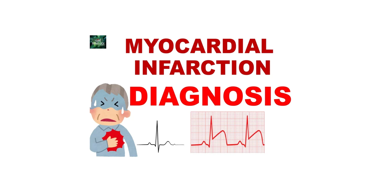
Electrocardiogram (ECG) changes are crucial for diagnosing myocardial infarction. A normal ECG includes a P wave, QRS complex, and T wave. The changes in ECG depend on whether the infarction is:
Transmural infarction:
Characterized by ST-segment elevation, referred to as ST elevation myocardial infarction (STEMI).
Subendocardial infarction:
Does not show ST-segment elevation.
Instead, may show ST-segment depression or T-wave inversion.
Referred to as non-ST elevation myocardial infarction (NSTEMI).
Watch this on youtube

