TRENDING:

Blood bags , types and uses
CHRONIC VENOUS CONGESTION – LUNG
Urine Analysis: Common Questions
Pathology Made Simple
  • Home
  • Education Contents
    • General Pathology
    • Systemic Pathology
    • Hematology
    • Clinical Pathology
    • Practical Pathology
      • Gross Pathology
  • Videos
  • Research
  • Pearls
  • Authors
  • About
    • Contact Me
    • Privacy Policy & Copyright notice
  • POSTGRADUATE
    • AUTOPSY

Select Page

Gross Pathology Specimen Series – Part 2

Posted by Dr Vijay Shankar S | Jul 1, 2025 | Practical Pathology, Gross Pathology |

Gross Pathology Specimen Series – Part 2

Welcome to Part 2 of the Gross Pathology Specimen Series — a visual learning resource for medical students and pathology enthusiasts. This series showcases classic and commonly encountered gross specimens, each accompanied by a short description to help you recognize key features, correlate with pathology, and prepare for practical exams or viva.

All images are optimized for fast viewing and learning, whether you’re revising on your laptop or mobile.


Carcinoma – Cecum

Ulceroproliferative mass distorting the cecal wall; firm, irregular, and malignant in appearance.
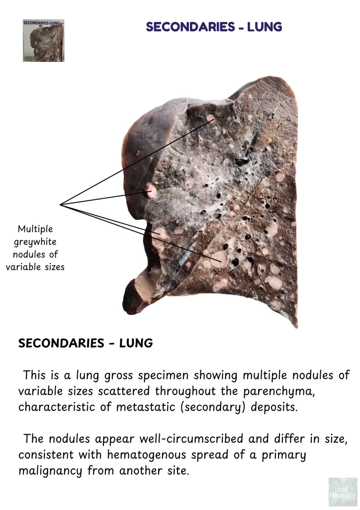
carcinoma cecum


Secondaries – Lung

Multiple grey-white nodules scattered across lung parenchyma, indicative of metastasis.

Secondary deposits - lung


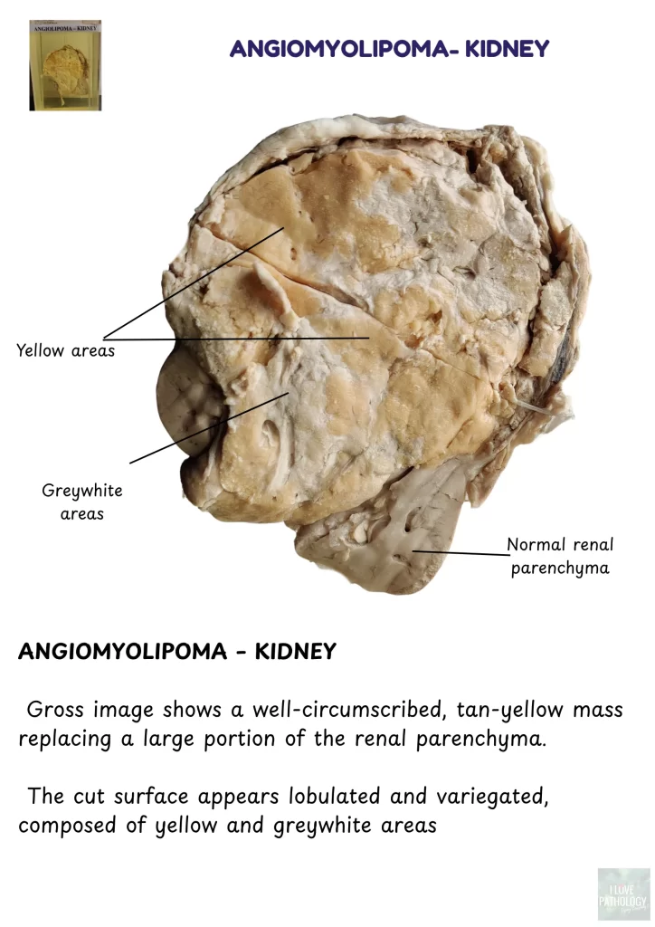
Angiomyolipoma – Kidney

Well-circumscribed yellow-grey lobulated mass replacing part of renal tissue.

angiomyolipoma kidny


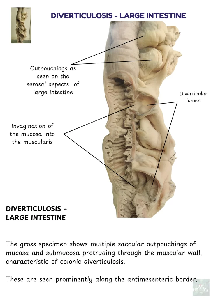
Diverticulosis – Large Intestine

Multiple saccular outpouchings of mucosa through muscle wall, along antimesenteric border.

diverticulosis


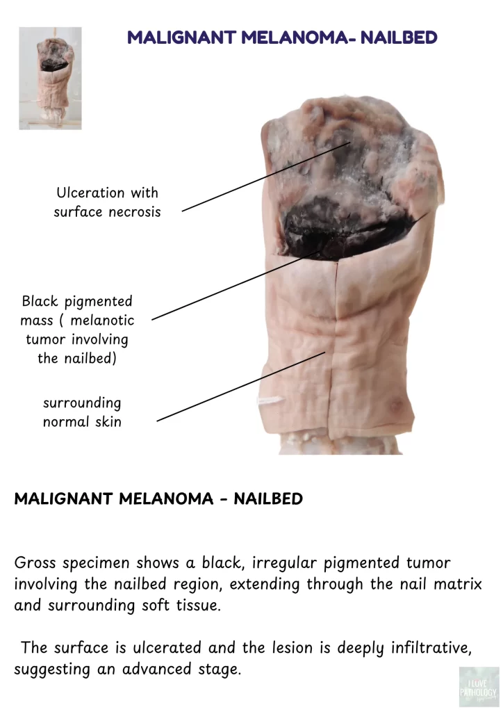
Malignant Melanoma – Nailbed

Black, irregular, ulcerated pigmented lesion extending through the nail matrix and soft tissue.
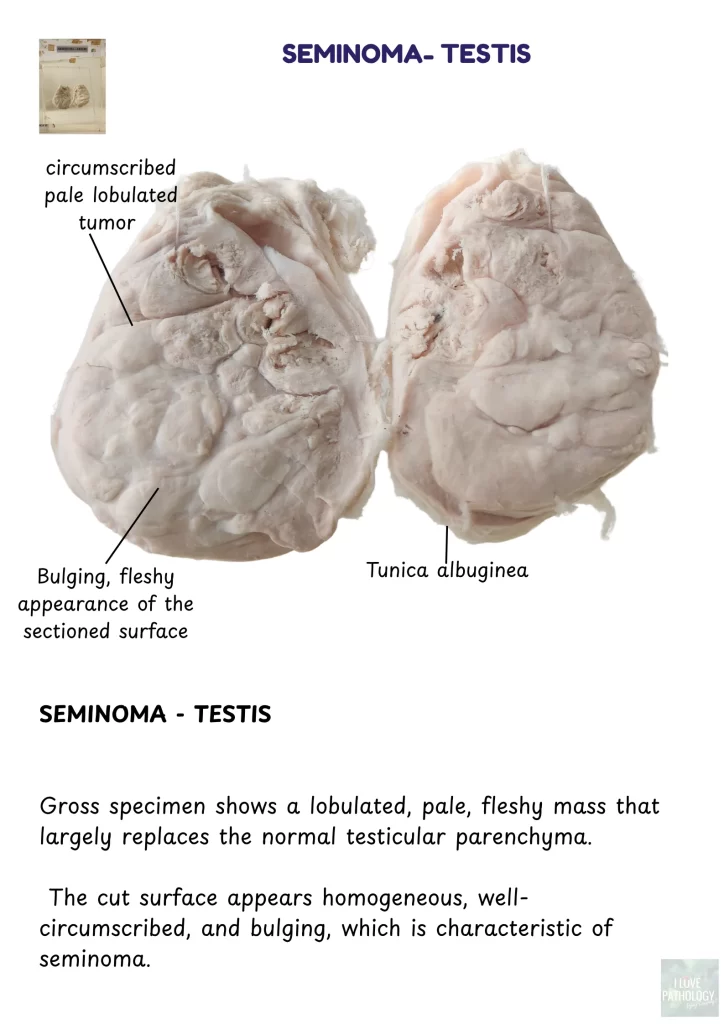
Malignant melanoma


Seminoma – Testis

Lobulated, pale, homogeneous, bulging tumor replacing normal testicular tissue.
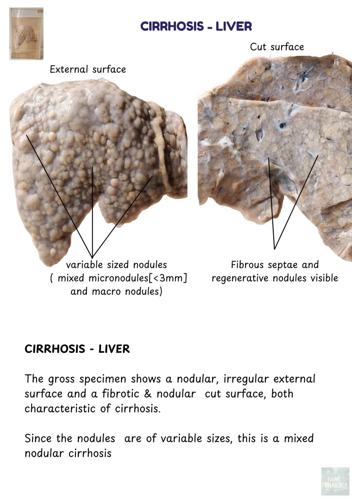
Seminoma


 Cirrhosis – Liver

Nodular, fibrotic liver with mixed micro and macronodules; distorted architecture.

Cirrhosis - liver


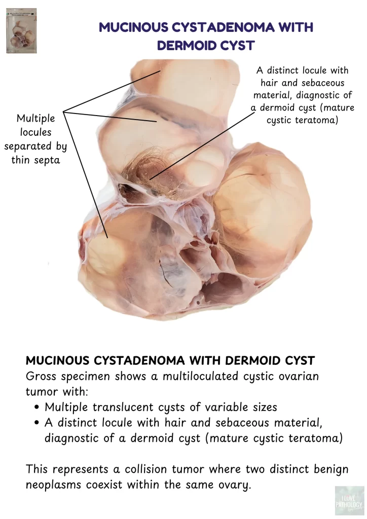
Mucinous Cystadenoma with Dermoid Cyst

Multiloculated ovarian tumor with translucent cysts and a distinct locule containing hair and sebum.

Mucinous cystadenoma with dermoid cyst


Share:

Rate:

PreviousWhat to Know Before Visiting a Cannabis Dispensary for the First Time
NextBenefits of Adding Mushroom Chocolate Bars to Your Routine

About The Author

Dr Vijay Shankar S

Dr Vijay Shankar S

With more than 20 years of experience as an academician, He has developed the art of content generation to make the learning more fun. Loves to promote social media for education in Pathology

Related Posts

LEPROSY- Tuberculoid & Lepromatous

LEPROSY- Tuberculoid & Lepromatous

May 15, 2021

Toxic Goiter

Toxic Goiter

May 19, 2016

Pathology Practical Illustrations : GENERAL PATHOLOGY

Pathology Practical Illustrations : GENERAL PATHOLOGY

November 3, 2023

Colloid Goiter

Colloid Goiter

May 19, 2016

Categories

ilovepathology

ilovepathology by Vijay Shankar

Flag Counter

Designed by Elegant Themes | Powered by WordPress