
Community Acquired Bacterial Pneumonia: Pathogenesis, Morphology, and Key Organisms
Community-acquired pneumonia (CAP) is a common acute infection of the lungs that affects otherwise healthy individuals who acquire the infection outside the hospital or healthcare setting. In this blog, we focus specifically on community-acquired bacterial pneumonia, its causative organisms, pathogenesis, and the morphological differences between lobar and bronchopneumonia.
Understanding Community-Acquired Pneumonia
CAP is broadly classified into:
-
Bacterial pneumonias
-
Viral pneumonias
Although clinical and radiological features often overlap between the two, one useful marker is procalcitonin, an acute phase reactant that is typically elevated in bacterial pneumonias.
Certain groups are at increased risk for developing CAP:
-
Extremes of age (very young or elderly)
-
People with chronic conditions such as congestive heart failure, COPD, or diabetes
-
Individuals with congenital or acquired immunodeficiencies
-
Those with decreased or absent splenic function (important for fighting encapsulated bacteria)
Why Splenic Function Matters
The spleen is a major source of anti-polysaccharide antibodies, which are essential in protecting against encapsulated organisms like Streptococcus pneumoniae. Impaired splenic function increases the risk of severe infections with such pathogens.
Common Causative Organisms in CAP
Let’s explore the key bacterial agents that cause community-acquired pneumonia:
1. Streptococcus pneumoniae
-
Gram-positive, lancet-shaped diplococcus
-
Most common cause of CAP
-
Still prevalent despite vaccination
-
Diagnosis: Presence of diplococci in neutrophils (may be false-positive due to normal flora)
2. Haemophilus influenzae
-
Gram-negative, pleomorphic bacillus
-
Encapsulated strains (especially Type B) are most virulent
-
Often follows viral respiratory infections
-
Important pediatric pathogen with high mortality
-
Also causes acute conjunctivitis (“pink eye”)
-
Most common bacterial cause of acute COPD exacerbations
3. Moraxella catarrhalis
-
Affects elderly, especially with cardiopulmonary conditions or diabetes
-
Second most common cause of acute COPD exacerbation after H. influenzae
4. Staphylococcus aureus
-
Follows viral illnesses like measles or influenza
-
High risk in IV drug users
-
Common complications: lung abscesses, empyema
5. Klebsiella pneumoniae
-
Common in chronic alcoholics and debilitated individuals
-
Characteristic feature: Thick, mucoid, blood-tinged sputum
-
Difficult to expectorate due to viscosity
6. Legionella pneumophila
-
Causes Legionnaires’ disease (epidemic/sporadic pneumonia) and Pontiac fever (self-limiting URI)
-
Found in water cooling systems, showerheads, hot tubs
-
Transmitted via inhalation or aspiration of contaminated water
-
High risk in transplant recipients or those with cardiac, renal, or hematologic conditions
-
Diagnosis: Detection of urinary antigen or sputum PCR
Morphological Patterns: Lobar vs Bronchopneumonia
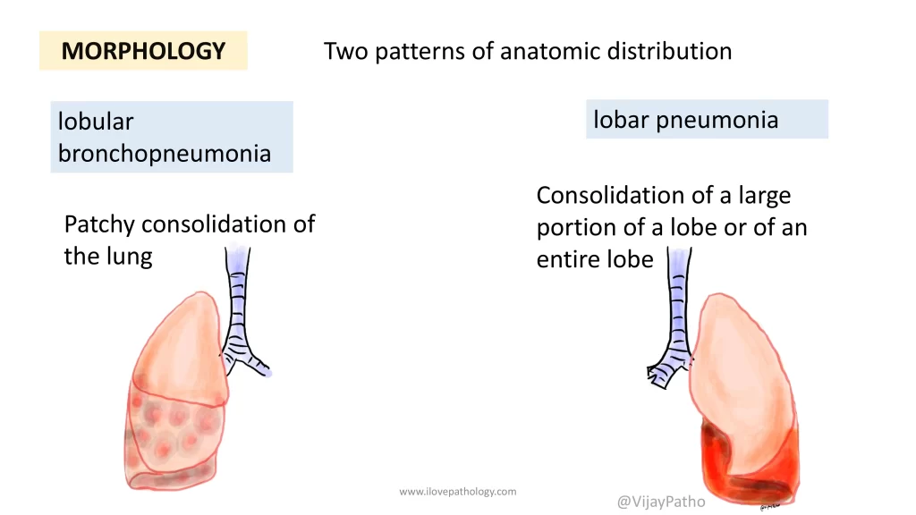
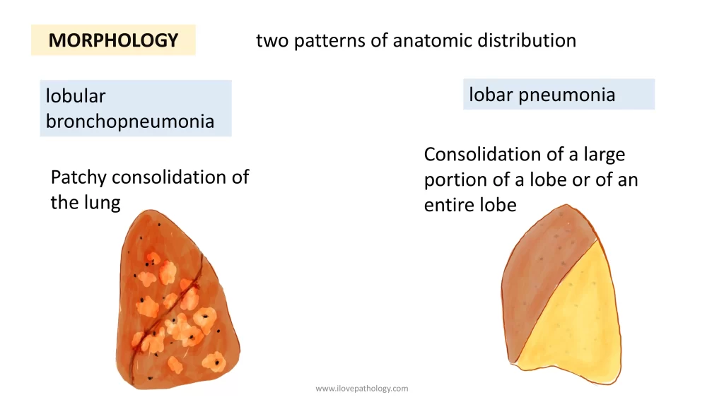
Consolidation is a key term—it means solidification of the normally air-filled lung tissue, due to the alveoli being filled with inflammatory exudate.
Bronchopneumonia (Lobular Pneumonia)
-
Patchy consolidation, often at the lung bases
-
Can be bilateral and multilobar
-
Lesions are gray-red to yellow, slightly elevated, dry, and granular
-
Microscopically: Suppurative inflammation in bronchi, bronchioles, and adjacent alveoli
-
Intervening lung tissue appears normal and aerated
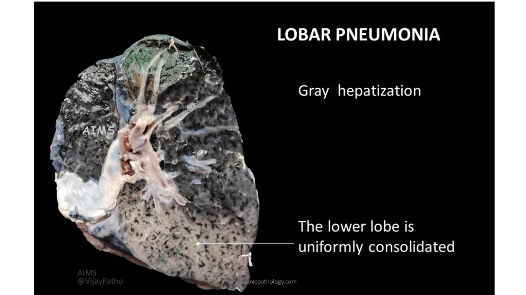
Lobar Pneumonia
-
Involves a large portion or the entire lobe
-
More uniform consolidation than bronchopneumonia
-
Common in healthy adults
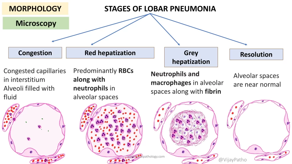
Classic Four Stages of Lobar Pneumonia:
-
Congestion
-
Lung appears red, heavy, boggy
-
Vascular engorgement with proteinaceous exudate in alveoli
-
-
Red Hepatization
-
Lung becomes firm, red, and liver-like
-
Alveoli filled with red blood cells, neutrophils, and fibrin
-
-
Gray Hepatization
-
RBCs disintegrate, lung appears gray-brown
-
More macrophages and fibrin deposition
-
-
Resolution
-
Exudate is cleared by macrophages or coughed out
-
Lung architecture returns to normal
-
Note: Due to early antibiotic treatment, it is rare to observe all these stages in clinical practice today.
Clinical Features of CAP
-
Abrupt onset of high-grade fever, shaking chills, and cough
-
Muco-purulent sputum, sometimes blood-tinged (hemoptysis)
-
Pleuritic chest pain and pleural rub if pleura is involved
-
These symptoms are due to the inflammatory response—especially interleukin-1 from immune cells
Diagnosis and Treatment
-
Chest X-ray helps identify the pattern:
-
Lobar pneumonia shows homogeneous opacity of one lobe
-
Bronchopneumonia shows patchy opacities
-
-
Definitive diagnosis: Culture of the pathogen and antibiotic sensitivity testing
-
Treatment: Appropriate antibiotics based on suspected/confirmed organism
-
Clinical improvement is usually seen within 48–72 hours
-
<10% of patients may require hospitalization
Complications (If Not Treated Properly)
-
Lung Abscess
-
Due to tissue destruction and necrosis
-
Common with Streptococcus pneumoniae and Klebsiella
-
-
Empyema
-
Pus in the pleural cavity
-
Involves fibrinous and purulent reaction in pleura
-
-
Bacteremic Dissemination
-
Spread of bacteria via blood to other organs
-
Can result in:
-
Myocarditis (heart)
-
Meningitis (brain)
-
Renal abscess
-
Suppurative arthritis (joints)
-
-