SECONDARIES / METASTATIC DEPOSITS IN LUNG
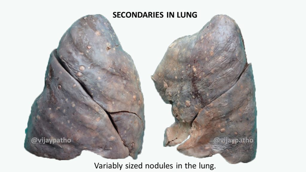
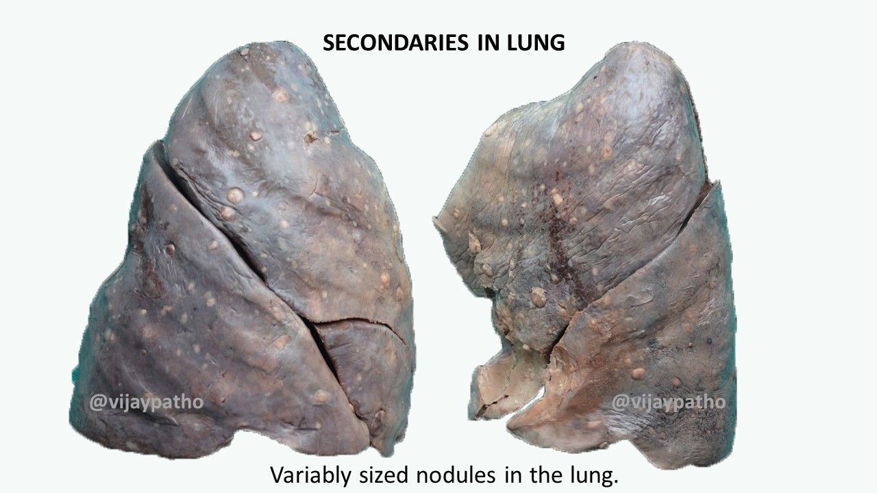
Multiple nodules in the lung parenchyma which are greater than one cm in diameter are most often metastatic deposits from a solid tumor.
The nodules are of varying sizes, discrete and are usually spherical but can be of various shapes too with irregular edges. Secondaries in lung/ metastatic deposits in lung
Can one differentiate probable primary tumor form the gross appearance?.
Yes . To a certain extent.
In a lobectomy specimens/ specimens from autopsy if one finds multiple nodules, based on the gross features, the probable site of origin can be guessed.
For example
BASED ON COLOR
Golden yellow: Renal cell carcinoma, sex cord stromal tumors, adrenal cortical tumors etc
Black/dark brown: melanoma
Red/golden brown: vascular tumors like angiosarcoma/kaposis sarcoma
BASED ON CONSISTENCY
Soft Hemorraghic with solid & cystic areas: Germ cell tumors
Firm to hard: Mesenchymal tumors like osteosarcoma, or any matrix producing tumors
Fish flesh like: lymphomas
Mucoid/ glistening: mucin secreting adenocarcinoma or other myxoid soft tissue tumors
MICROSCOPY
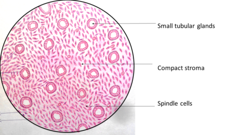
Most of the metastatic deposits on microscopy resembles that of the primary tumor
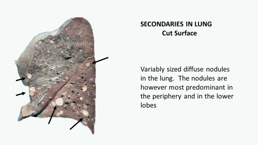
What is miliary metastasis?
Metastatic deposits in the lung presenting as variably sized diffuse nodules in the lung. The nodules are however most predominant in the periphery and in the lower lobes
the most common carcinomas presenting as miliary metastasis to lung are
thyroid, breast , renal cell carcinoma, colorectal, melanoma etc.
Other tumors include osteosarcoma, trophoblastic tumors, pancreatic tumors etc.
What are the infective causes of miliary nodules in the lung?
The causes include
Tuberculosis
Dissimenated fungal diseases like histoplasmosis and blastomycosis
Viral infections like cytomegalovirus infections in immunocompromised patients.
What are the mechanisms of metastasis to lung?
Hematogenous spread
Lymphatic spread
Direct extension of a malignancy of a contiguous structure like , trachea, esophagus, thyroid, thymus and chest wall

