
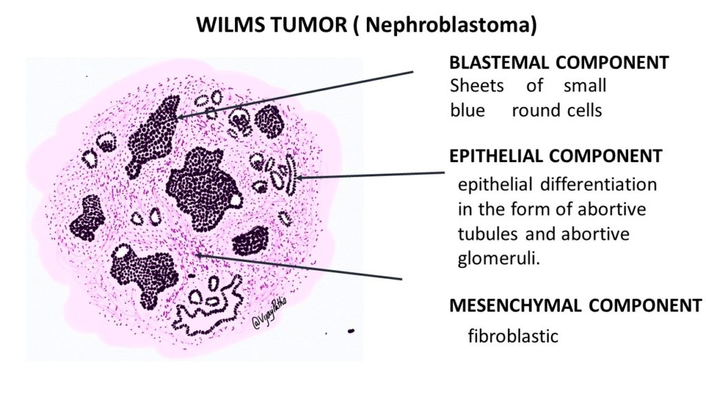
WILMS TUMOR (NEPHROBLASTOMA)
It is the most common pediatric renal tumor.Occurs in childern between 2- 5 years of age.
Majority of the tumors are sporadic.
But can also be associated with these syndromes namely
1. WAGR syndrome ( Wilms tumor, Aniridia, Genital abnormalities, and
mental Retardation)[ deletion of WT1 gene]
2. Denys- Drash Syndrome ( gonadal dysgenesis and early onset nephropathy )[negative inactivating mutation in WT1]
3. Beckwith-Wiedemann syndrome ( associated with hemihypertrophy, hepatomegaly, renomagaly, macroglossia, adrenal cytomegaly)[ due to genomic imprinting principally involving the IGF2 gene.]
Morphology:
Gross: Large well circumscribed mass. Can be multicentric and bilatral in small percentage of cases. on cut section, the tumor is soft, homogenous greywhite to tan. Hemorrhage necrosis and cystic degeneration can be seen.
Microscopy:
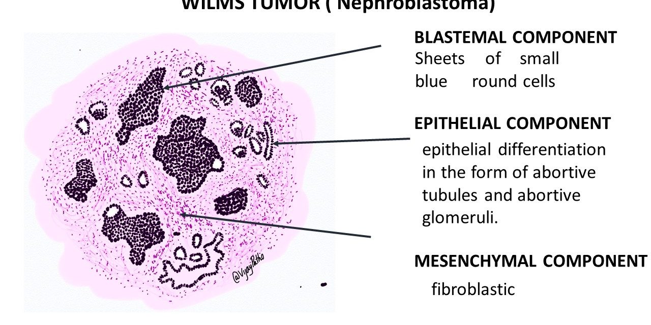
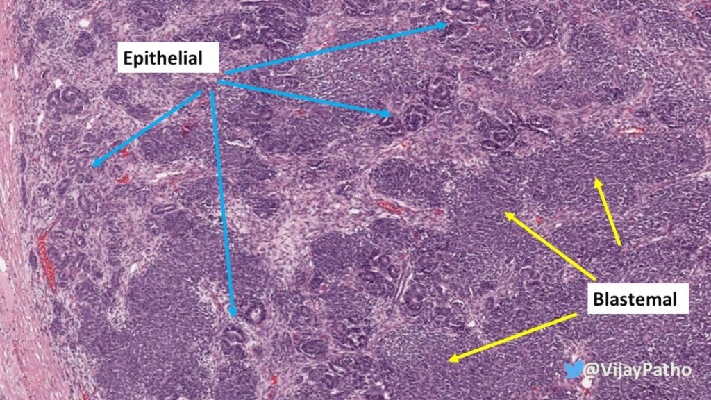
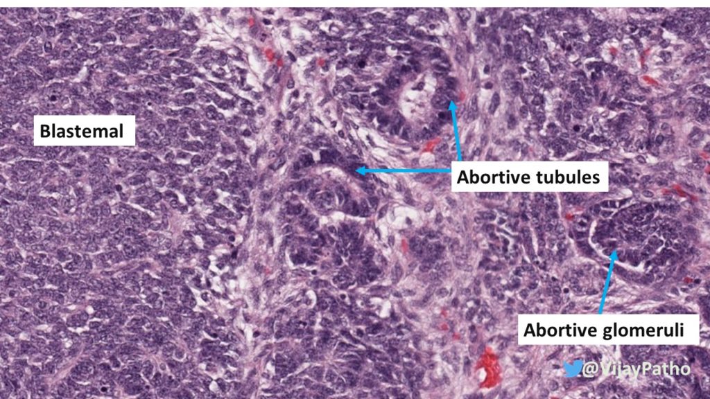
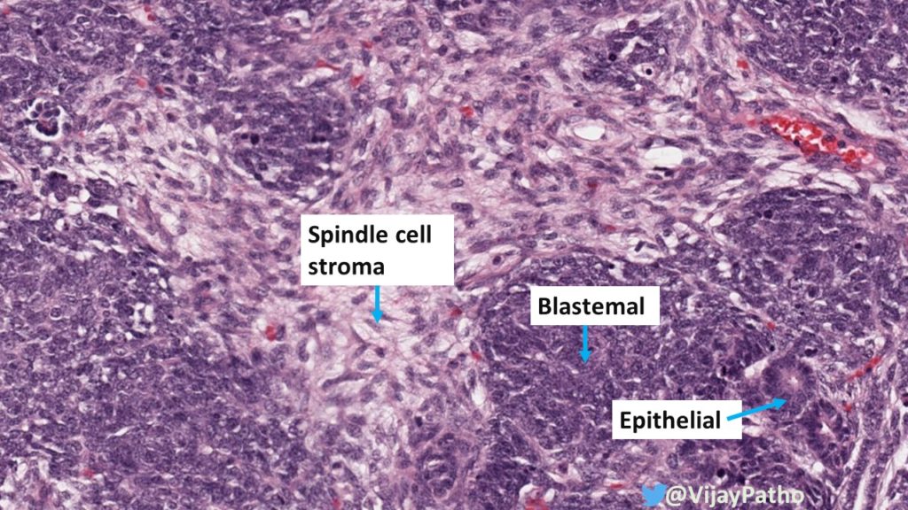
Classical histopathological features is the triphasic pattern comprised of Blastemal, epithelial and stromal components in variable proportions
Blastemal component: Sheets of small blue round cells. This is the least differentiated cellular element.
Epithelial component: These cells resemble those of epithelial differentiation in the form of abortive tubules and abortive glomeruli. The tubules can be primitive rosette like tubules or may be well formed tubules.
Stromal component: can be fibroblastic or myxoid. May show heterologous elements like skeletal muscle, adipose tissue , smooth muscle, bone , cartilage or even a neuroglial tissue.
Sometimes Wilms tumor contain only one or two of the elements, when they are referred to as monophasic and biphasic respectively.
Anaplasia can be present but it is often a marker of unfavorable prognosis.
the illustrated image of WILMS tumor is a below
Visit pathpresenter.net for amazing collection of slides for learning histopathology.


Recent Comments