
Dear All
The Part 9 of ” Similes & Metaphors of Pathology” is here. ENJOY LEARNING 🙂
Note: Mobile users please use landscape mode for complete view.
More interesting ones to come in Part 10!….
Comments and suggestions are welcome.
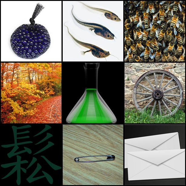
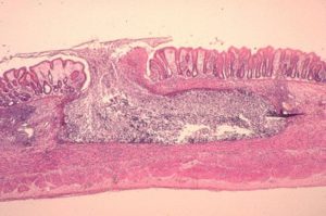
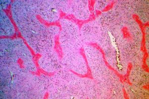
Bag of Marbles |
Bag of marbles appearance in Myospherulosis ( source ) |
Bag of marbles appearance in Myospherulosis
Myospherulosis is an uncommon foreign body reaction that occurs in tissues exposed to antibiotic and oil-based ointments. Characterised by the presence of sac like structures containing spherules which give the appearance of partly filled bag of marbles. It is not a fungal infection |
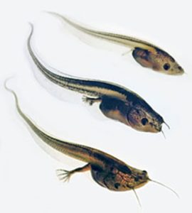
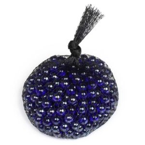
Tadpoles |
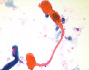
Tadpole cells in squamous cell carcinoma( source ) |
Tadpole cells in squamous cell carcinoma these cells are Elongated and club shaped. These cells have broad end which tapers to narrow end and has hyperchromatic nucleus with dense keratinized cytoplasm, which looks like a tadpole.
sometimes also seen in Rhabdomyoblastoma, where these cells are called as strap cells/tadpole cells. |
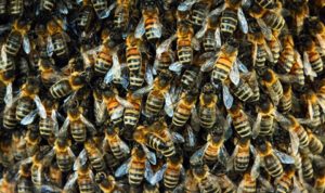
Swarm of bees |
 swarn of bees ( source ) |
Swarm: a group of honey bee that emigrate from hive and fly off to start a new colony OR bees settled in a hive.
Swarm of bees in alopecia areata is used to desctibe hair bulbs in alopecia areata which are diffusely infiltrated by discrete clusters of lymphocytes. Also referred to as lymphocytes buzzing in peribulbar region appearance. This is seen in Acute stage of Alopecia areata.
|

Vermont Foliage |
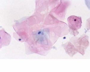
Vermont foliage appeareance of hyperkeratosis ( source ) |
Vermont is a state in the New England region of the northeastern United State.
Foliage means fall of leaves/cluster of leaves Vermont foliage appeareance of hyperkeratosis in cervicovaginal smears of pap smears. Presence of numerous anucleate appearance in pap smears of hyperkeratotic lesion, resembles that of foliage in vermont. An uncommonly used simile! |
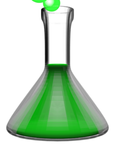
Flask |
Flask-shaped ulcer of intestinal amebiasis.( source ) |
Flask-shaped ulcer of intestinal amebiasis.
Caused by E histolytica, Amebic lesions begin as small foci of necrosis that progress to ulcers. Muscle coat of the large intestine form a barrier to the penetrating trophozoites and hence, the ulcer fan out latarally resembling flask-shaped ,with narrow neck and broad base |
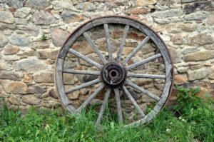
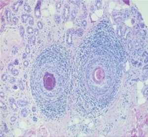
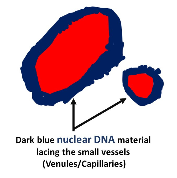
Cart wheel |
Cart wheel nucleus of Plasma cells.( source ) |
Cart wheel nucleus of Plasma cells.
Plasma cells are large lymphocytes with charcterestic featires They have basophilic cytoplasm and an eccentric nucleus. The nucleus with heterochromatin is in a characteristic cartwheel or clock face arrangement. There is a perinulear halo, that on electron microscopy contains an extensive Golgi apparatus and centrioles |

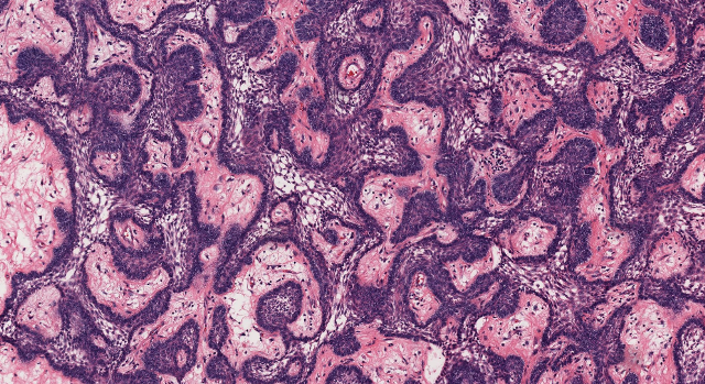
Chinese letters |
Chinese letter pattern in Fibrous dysplasia ( source ) |
Chinese letter pattern in Fibrous dysplasia
Fibrous dysplasia (FD) of bone is an uncommon disease caused by sporadic, congenital mutations in the cAMP regulating protein, Gsα. classic “Chinese characters” on histopathology is made of trabeculae of bone with fibrous tissue interwoven between |
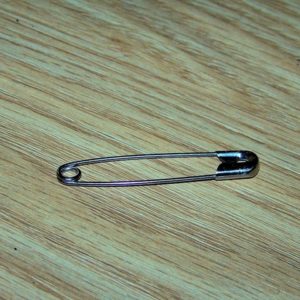
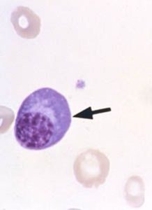
Safety pin |
Safety-pin’ appearance of Klebsiella granulomatis ( source ) |
Safety-pin’ appearance of Klebsiella granulomatis causing Donovanosis or granuloma inguinale
The organisms are seen within the cytoplasm of histiocytes from the smear taken from the ulcer.. The bacteria are ovoid or bean-shaped varying in size from 1 to 1.5 pm in length and 0.5 to 0.7 pm in thickness. When stained by Giemsa, they have blue chromatin inclusions at each pole/bipolar staining which gives the characteristic ‘closed safety-pin’ appearance. |

Envelope |
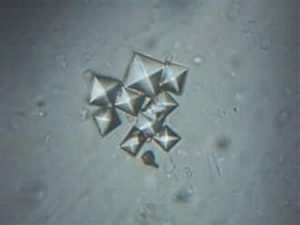
“envelope”) shaped crystals of calcium oxalate ( source ) |
octahedral (“envelope”) shaped crystals of calcium oxalate
Appearance Dihydrate: octahedral (“envelope”) Monohydrate: dumbbell, ovoid, or rectangular in shape. Major component of renal calculi. |


Recent Comments