
Rosettes in Pathology:
Rosettes are structures with radial arrangement of cells like that of a rose petals and hence the name “rosette”. Presence of these structures are diagnostic of certain tumors.
There are different kinds of rosettes we encounter in pathology. These are as described below
Homer Wright Rosettes: Here the rosettes have a dense feltwork of interwoven cytoplasmic processes of nerve cells and neuroglial cells( Neuropil) in the centre. This is named after James Homer Wright an American pathologist(1869 – 1928 )
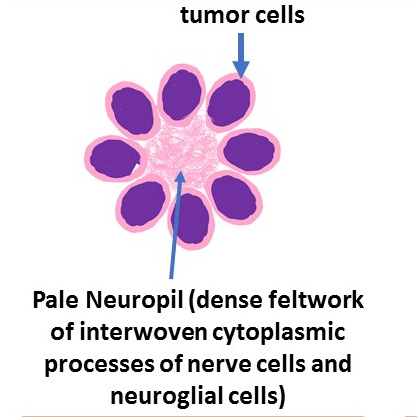
These rosettes are seen in
Neuroblastoma
Medulloblastoma
Pineoblastoma
Flexner-Wintersteiner Rosette: First described by first described by Simon Flexner an American Physician and Pathologist and Austrian ophthalmologist Hugo Wintersteiner. Characteristicaklly seen in retinoblastomas. Here the rosette surround a central lumen containing small cytoplasmic extensions of the encircling tumor cells. unlike Homer Wright rosette, the lumen do not have Neuropil. The tumor cells forming the Flexner–Wintersteiner rosette have features of primitive photoreceptor cells when examined ultrastructurally.
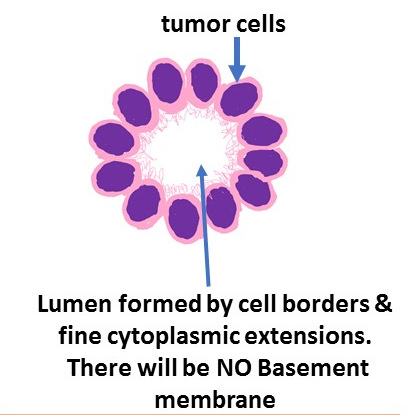
The other tumors in which these are seen are
pinealobastoma
Medulloepithelioma
Perivascular Pseudo Rosettes : These are called “pseudo” these are not true rosettes and the tumor cells surrounds a blood vessel and hence “perivascular” These are charactestically seen in ependymomas. In this type of rosette, the neuropil is seen projecting towards a central blood vessel.
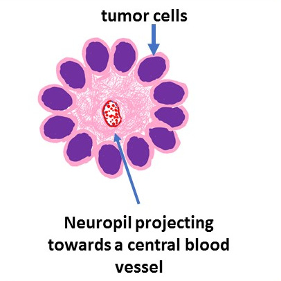
The other tumor in which these are seen are
Central Neurocytoma
Glioblastoma


Recent Comments