
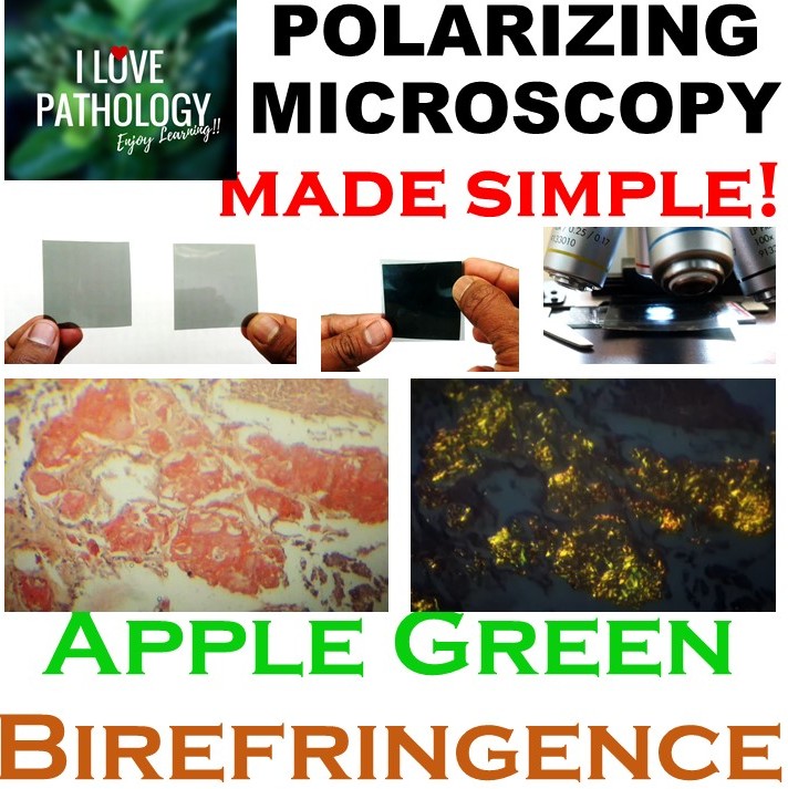
Polarizing microscopy: It is a type of Optical microscopy which uses polarized light for examination of specimens/slides.
This type of microscopy was developed initially to examine crystalline structures found in rocks and minerals. However, this is also of use in the field of medicine.
The application of polarizing microscopy in the field of Medicine Includes
- Examination of urine sediment
- Gout
- Demonstration of Amyloid etc..
What we need to understand is that we don’t have to own a dedicated polarizing microscope to examine these specimens. Just a pair of polarizing filters are enough which can be effectively used with an ordinary binocular microscope.
Polarizing filters: These are the filter sheets which can be available online and in scientific stores. You need two of them.
These two filters if they are not crossed, they allow light to enter. If they are crossed they will not allow light to enter through them. It is through this “crossed position” the specimens are observed.
Procedure :
- Place one filter above the light source. This acts as “polarizer”
- The second filter should be placed on the slide. This acts as analyzer.
- Use 10X objective to examine slides. Higher powers cannot be used effectively with the presence of these sheets in between the slides and the eye piece.
- Gently rotate the “polarizer” on the light source as you are viewing through the eye piece so that it is in “crossed position”. At this position you can see brilliant Birefringence of the specimens/material on the slide.
The above video shows the brilliant “Apple Green Birefringence” of the amyloid which is stained with Congo red stain. The slide used was medullary carcinoma of thyroid.
We all know that Medullary carcinoma of thyroid is a type of thyroid carcinoma which originates from parafollicular cells/C cells. These secrete calcitonin. One of the characteristic feature is the presence of amyloid in the stroma of these tumors.
Amyloid as we all know is an extracellular, fibrillary insoluble proteinaceous substance. On Spectroscopy these amyloid fibrils demonstrate cross beta pleated sheet configuration. It is this cross beta pleated configuration, which is important for the staining characteristics of amyloid with congo red ( the amyloid is stained pink/red) as well as its characteristic “Apple green Birefringence” on polarized microscopy. This demonstration is more specific in diagnosis of amyloidosis.
Isn’t this technique so simple to demonstrate one of the most beautiful characteristics of Amyloid?
Enjoy learning!


Recent Comments