
PLEXIFORM PATTERN #patternsinhistopathology
The word ‘plexiform’ denotes something which resembles or forms a plexus or a network.
Plexus means a branching vessels or nerves. Eg Brachial plexus which is a nerve plexus formed by intercommunications among the ventral rami (roots) of the lower 4 cervical nerves (C5-C8) and the first thoracic nerve (T1), as shown in the illustration below
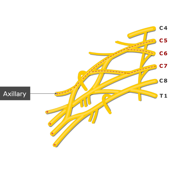
Brachial plexus
Plexiform pattern in histopathology denotes a histomorphological pattern which shows a network of interconnecting strands of cells. It also denotes a coplex multinodular lesion.
Plexiform pattern predominantly noted in neural tumors like neurofibroma, schwannoma perineurioma, granular cell tumors or sometimes in malignant peripheral nerve sheath tumors.
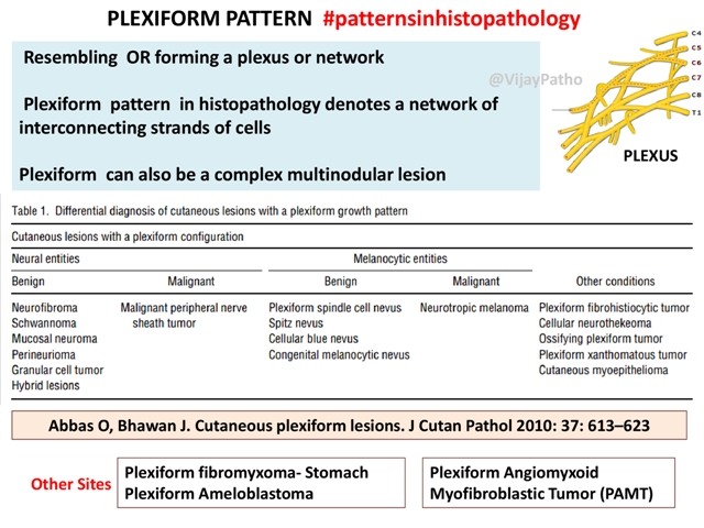
Some of the types of melanocytic nevus like spitz nevus, cellular blue nevus or a plexiform spindle cell nevus can show a plexiform pattern. Rarely malignant melanoma can also show a plexiform pattern.
Other conditions where plexiform pattern is noted are as below
• Plexiform ameloblastoma
• Plexiform fibromyxoma
• Plexiform fibromyxoid myofibroblastic tumor(PAMT)
• Plexiform fibrohistiocytic tumor
• Cellular neurothekeoma
You can go through this article which encompasses various cutaneous plexiform lesions:
Abbas O, Bhawan J. Cutaneous plexiform lesions. J Cutan Pathol 2010: 37: 613–623.
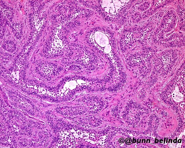
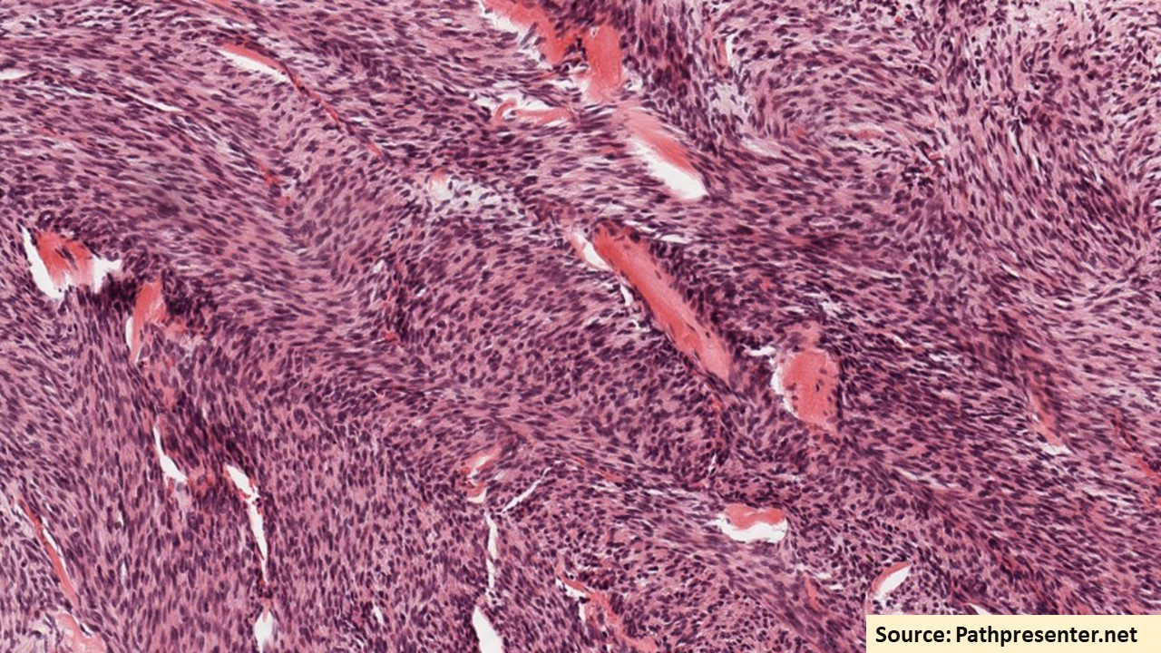
The above image is a case of ameloblastoma with beautiful plexiform growth pattern. This and other beautiful images shared by Belinda Bunn @bunn_belinda on twitter can be accessed from the link below
https://twitter.com/bunn_belinda/status/874812283314216961
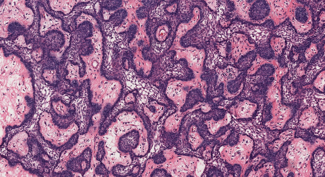
This is another beautiful example of plexiform ameloblastoma showing anastomosing trabeculae and cords of epithelial cells. This slide can be accessed as a virtual slide at pathpresenter.net in the link below.
More to follow…


Recent Comments