
OSTEOSARCOMA
Osteosarcoma is the second most common primary malignant skeletal neoplasm and accounts for approximately 20% of all primary malignant bone tumors. The most common bone tumor is myeloma.
Osteosarcoma is defined as a malignant tumor in which the bone matrix is produced by tumor cells.
long bones are more commonly involved., although any bone can be involved.
Some of the conditions are associated with osteosarcoma. These include
1. Pagets disease
2. Chronic osteomyelitis
3. Post irradiatin
4. Infarction of bone
5. Prosthetic implants
6. Retinoblastoma
Osteosarcoma has Bimodal age distribution with majority occurring before age 25, and rest around 20-30% occurring in individuals older than 40 years.
Males are more commonly affected than females.
Classification:
Based on anatomical site, they are broadly classified into intramedullary(classical/conventional) or surface osteosarcoma. Both these types can be low grade or high grade tumors. Rarely intracortical location is also seen.
Based on pathogenetic mechanism, the are categorized into primary and secondary( due to preexisting /associated disorders as mentioned above)
These patients present with localized pain with or without mass/swelling. Pathologic fracture may be the initial manifestation in few cases of osteosarcoma.
It is important to know that low grade osteosarcomas usually presents as painless mass forming lesion.
Radiologically, these are large destructive lesions which are radiodense lesions due to the deposition of osteoid matrix. Two important findings are worth mentioning though the are not specific. They are “sunburst appearance” and “Codman’s triangle” ( as illustrated below)
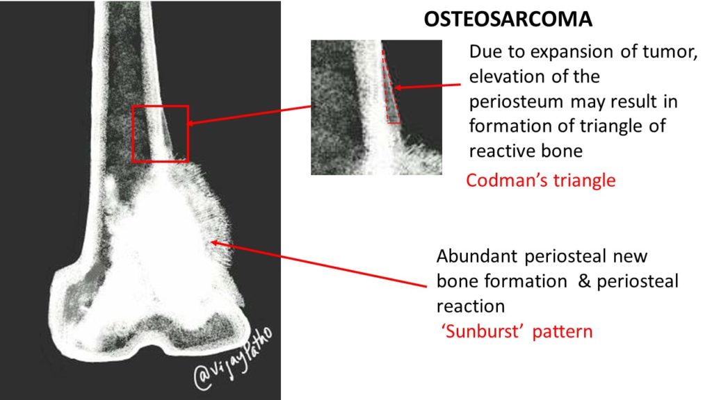
Cod man’s triangle, SUnburst pattern.. X ray findings of osteosarcoma
Gross: the morphology varies depending on the amount of bone present and other components. In majority of the cases, these are large and bulky tumors which are destructive, as it infiltrates into the surrounding soft tissue, and has variable hemorrhagic, necrotic and cystic areas.
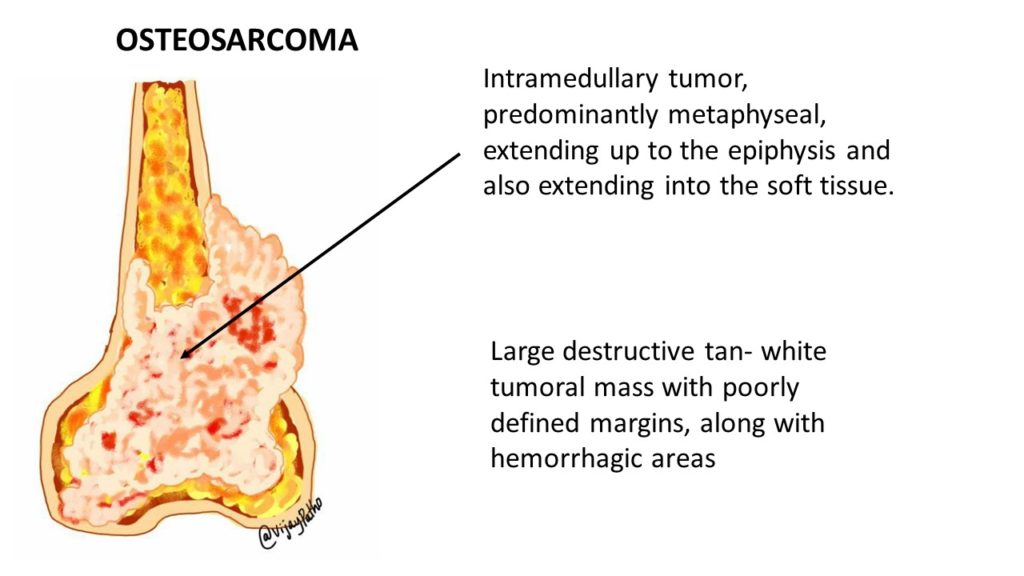
macroscopic appearance of osteosarcoma
Microscopy:
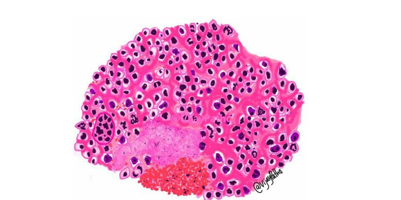
The tumor is composed of highly pleomorphic cells with hyperchromatic nuclei, prominent nucleoli , some of them with highly bizzare morphology. These tumor cells form woven bone which is neoplastic. This woven bone is primitive in nature and deposited as disorganized trabeculae around tumor cells, in a lace like pattern. This is referred to as Osteoid. Sometimes, this bone can be in the form of broad sheets. Numerous mitosis is common. Areas of hemorrhage and necrosis is often seen.
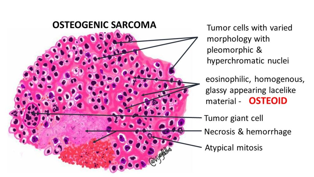
illustrated pathology of osteosarcoma
Note that the microscopic appearance is highly variable and depends on the type of osteosarcoma.
Histologically there are different types , depending on the predominant type of matrix. These are
a. Osteoblastic
b. Chondroblastic
c. Fibroblastic
d. Small cell
e. Telangiectatic
CLICK HERE to the virtual slide of ostrosarcoma.
Want to know about another important bone tumor?
CLICK HERE to learn about Giant cell tumor of bone.


Recent Comments