

Let us understand some basic concept of cytoplasmic and nuclear changes in irreversible cell injury
CYTOPLASMIC CHANGES:
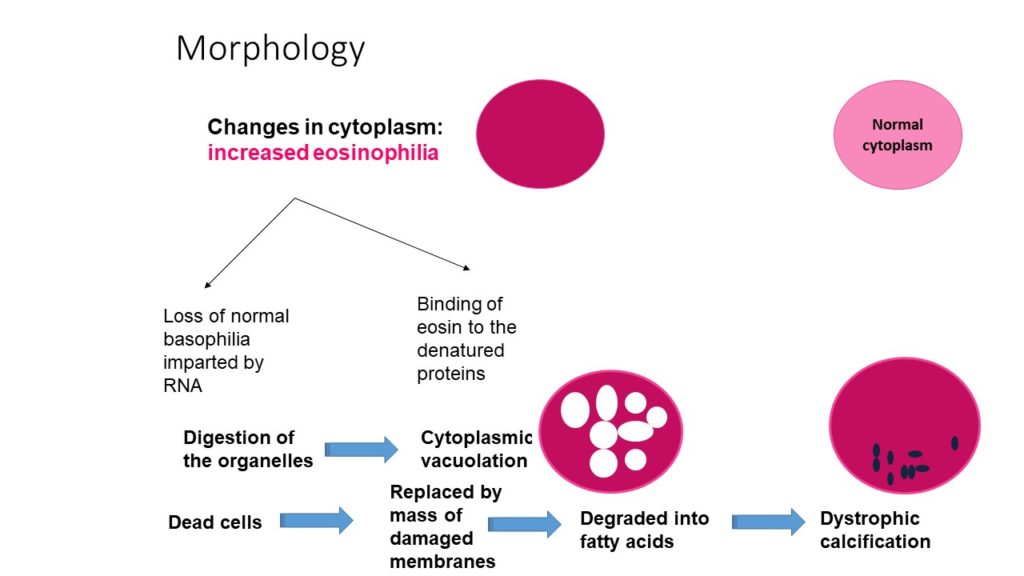
1. Looks more pink! INCREASED EOSINOPHILIA:
Two important reasons for this
a. In a routine Hematoxylin and Eosin staining of a tissue, the cytoplasm of the cells though stains pink, there is some amount of basophilic hue due to the presence of RNA in the cytoplasm. Now, during the process of irreversible injury, the RNA will be digested by activation of RNAses ! SO, The basophilic Hue imparted by RNA is lost and hence the cytoplasm looks relatively more PINK!
b. The cell injury also means, the cytoplasmic proteins are denatured. The dye Eosin, in H & E stain, binds to the denatured proteins and hence the cytoplasm looks more PINK!
2. Over a period of time, the organelles are also digested resulting in vacuolations in the cytoplasm. Another important change is CYTOPLASMIC VACUOLATION
3. DYSTROPHIC CALCIFICATION can also occur, when the mass of damaged membranes are degraded into fatty acids.
NUCLEAR CHANGES
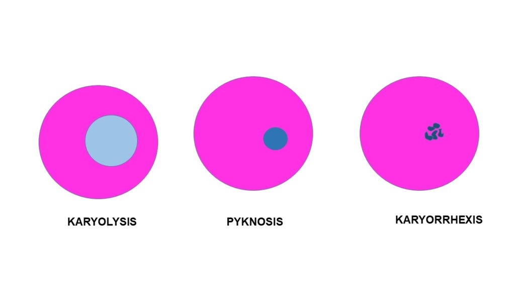
These changes occur due to nonspecific changes in the DNA as a result of DNAse activity. Remember, DNAses are activated in irreversible cell injury.
The changes are
1. KARYOLYSIS: This is essentially because of DNAse activity. There is dissolution of nuclear DNA, which result in loss of affinity of nucleus to nuclear stain. Hence the nucleus fades, because, normally the basophilia in nucleus is because of the chromatin. When the chromatin disintegrates, the basophilia also fades.
2. PYKNOSIS: As the chromatin is disintegrated, there is condensation of DNA into a small mass resulting in shrinkage of nucleus. And as a result of shrinkage and condensation , then mass appear more darker. This is PYKNOTOC nucleus.
3. KARYORRHEXIS: The pyknotic nucleus also eventually get fragments and is referred to as KARYORRHEXIS.
Finally the nucleli disaapears!
If you want to hear the explanation in detail CLICK HERE to find the video on my Youtube channel


Recent Comments