
INTESTINAL TUBERCULOSIS
Tuberculosis affecting the intestines can be due to primary or secondary infection
- What is primary intestinal tuberculosis
This is the the tuberculosis of intestine caused by ingestion of unpasteurized milk which contains Mycobacterium bovis.
- What is secondary intestinal tuberculosis
This is a type of tuberculosis which occurs in a patient of active pulmonary tuberculosis who swallows the coughed up sputum and the lesions are developed in the intestine secondary to the swallowed material.
- What are the common sites of intestinal tuberculosis
The most common site of intestinal tuberculosis is ileum. Yet it can involve any part of the bowel from duodenum to rectum.
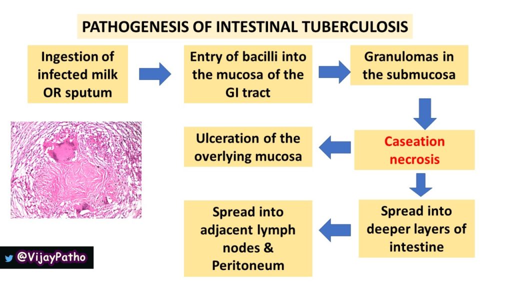
- Discuss the pathogenesis of Intestinal tuberculosis
The Pathogenesis of intestinal tuberculosis is explained in the illustration below.
- What are the different forms of intestinal tuberculosis
Intestinal tuberculosis can occur in three forms
a. Ulcerative ( most common) b. Hypertrophic/hyperplastic c. Ulcerohypertrophic
- Describe the morphological features of intestinal tuberculosis
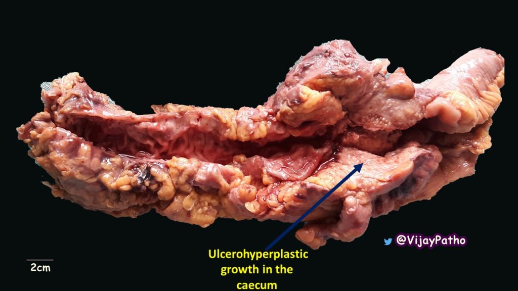
Gross Examination
The lesions in secondary intestinal tuberculosis begins as a small ulcerative lesion which later enlarges to form a large transverse ulcer i.e the ulcers are perpendicular to the long axis of the bowel. This is because of the spread by lymphatics. ( Note that this is in contrast to typhoid ulcers which are longitudinal ulcers, i.e, they run parallel to the long axis of the bowel, which is due to the involvement of Peyers patches )
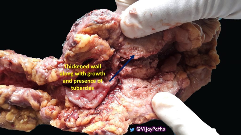
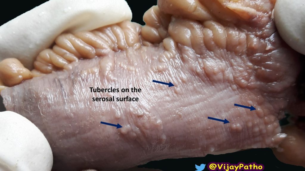
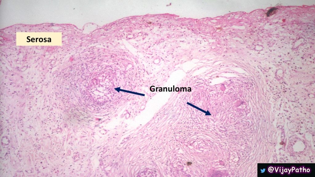
The serosal surfaces can show the presence of tubercles which is a small elevation or protuberance on the serosal surface. Later stages strictures of the small intestines are seen which is due to fibrosis. Long standing lesions may even show Napkin-ring line constriction !
Mesentric lymph nodes may be enlarged.
In Hyperplastic type of tuberculosis, the cecum is primarily involved, sometimes ascending colon. The wall of the cecum is thick and sometimes forms a mass lesion because of tuberculous granulation tissue . The mass can be palpable on clinical examination.
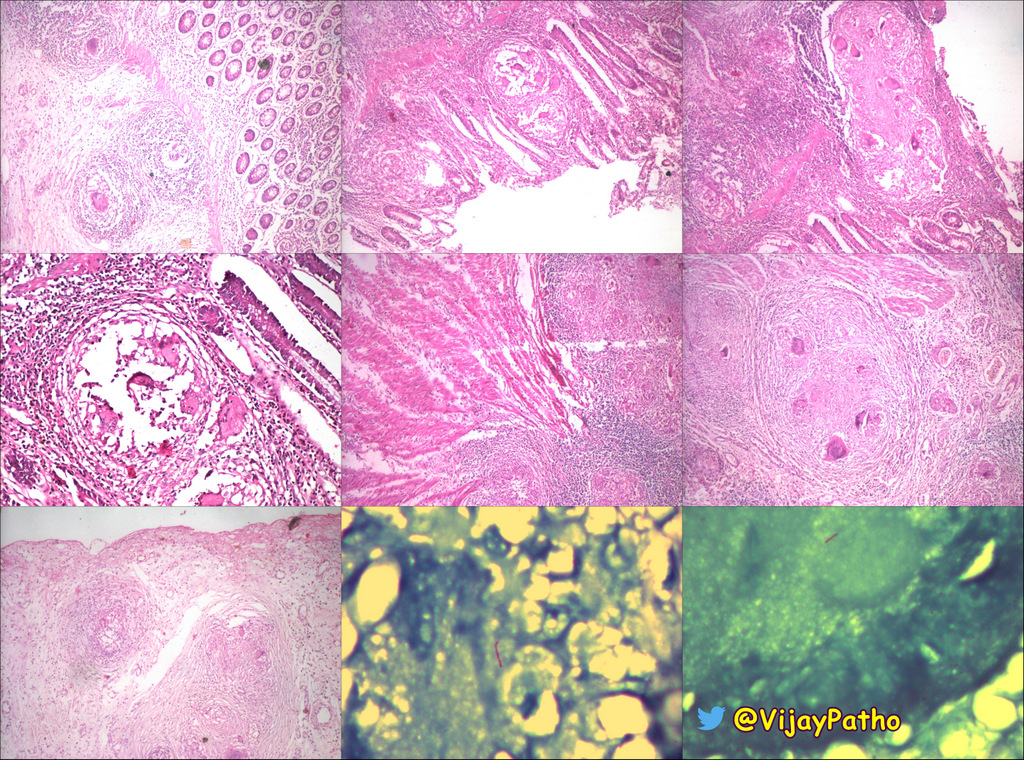
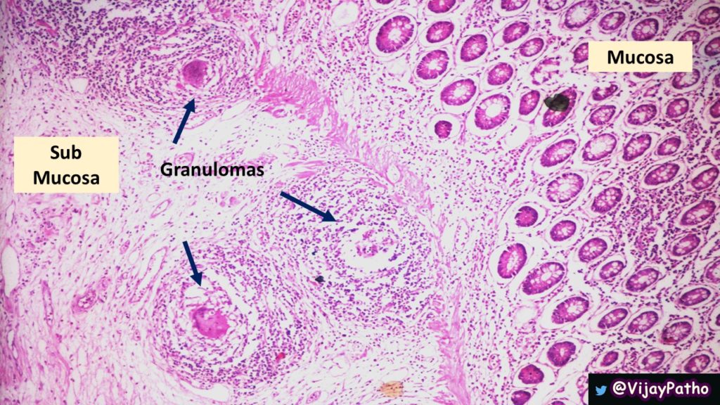
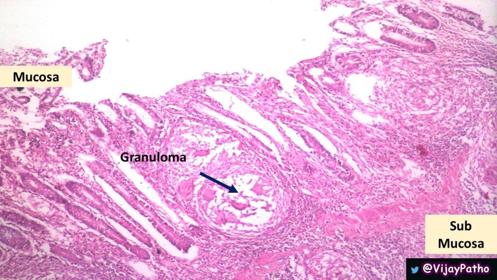
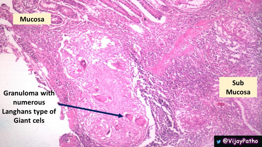
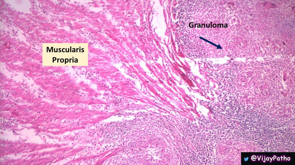
Microscopic examination
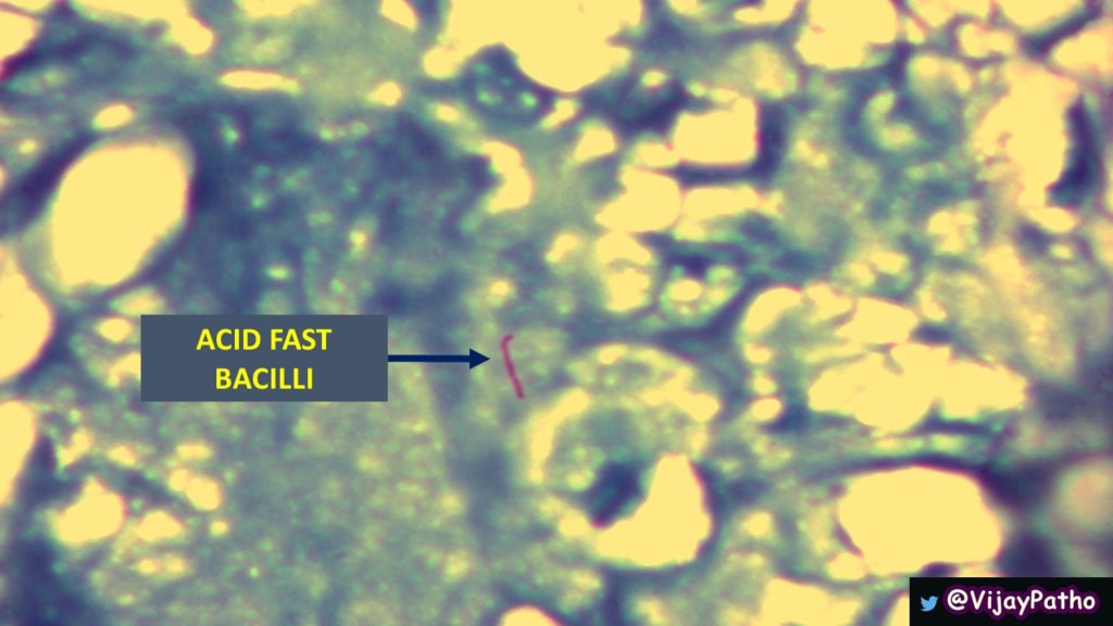
Mucosa shows ulceration. Caseating granulomas are seen in the wall of the intestine. The granulomas show marked variation in size. The periphery of the granulomas contain admixture of lymphocytes, plasma cells and Langhans type of giant cells. Sections stained for acid fast bacilli will be positive. Note that the sensitivity of AFB staining is very low (20%), yet a positive stain is 100% specific for intestinal tuberculosis.
- Mention the complications of intestinal tuberculosis
The complications include
- Tuberculous peritonitis
- Strictures due to fibrosis may result in intestinal obstruction.


Recent Comments