
Chronic Rheumatic heart disease
Acute rheumatic carditis may progress to chronic rheumatic heart disease.
“The myocarditis and pericarditis usually resolve without any sequelae. Valvular endocarditis may progress to the deforming fibrotic valvular visions which are the key feature of chronic rheumatic heart disease”
The mitral valve is the most commonly involved. The valve leaflets become thickened due to fibrosis.
“Fusion of the valve commissures results in a narrowed and fixed valvular opening which has a characteristic fish mouth or buttonhole appearance”
Shortening, thickening and fusion of the chordae tendinae occurs. The structural changes result in mitral stenosis with progressive dilatation of the left atrium, chronic venous congestion of the lungs, pulmonary hypertension, right ventricular hypertrophy and right sided heart failure.
The aortic valve is the second most commonly involved one. Thickening of the cusps and fusion of the commissures results in aortic stenosis.
The right sided valves ie. the tricuspid and pulmonary valves are seldom involved.
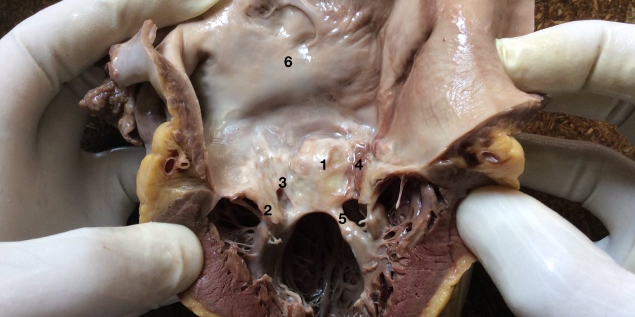
The photograph shows the left atrium, left ventricle and the stenosed mitral valve.
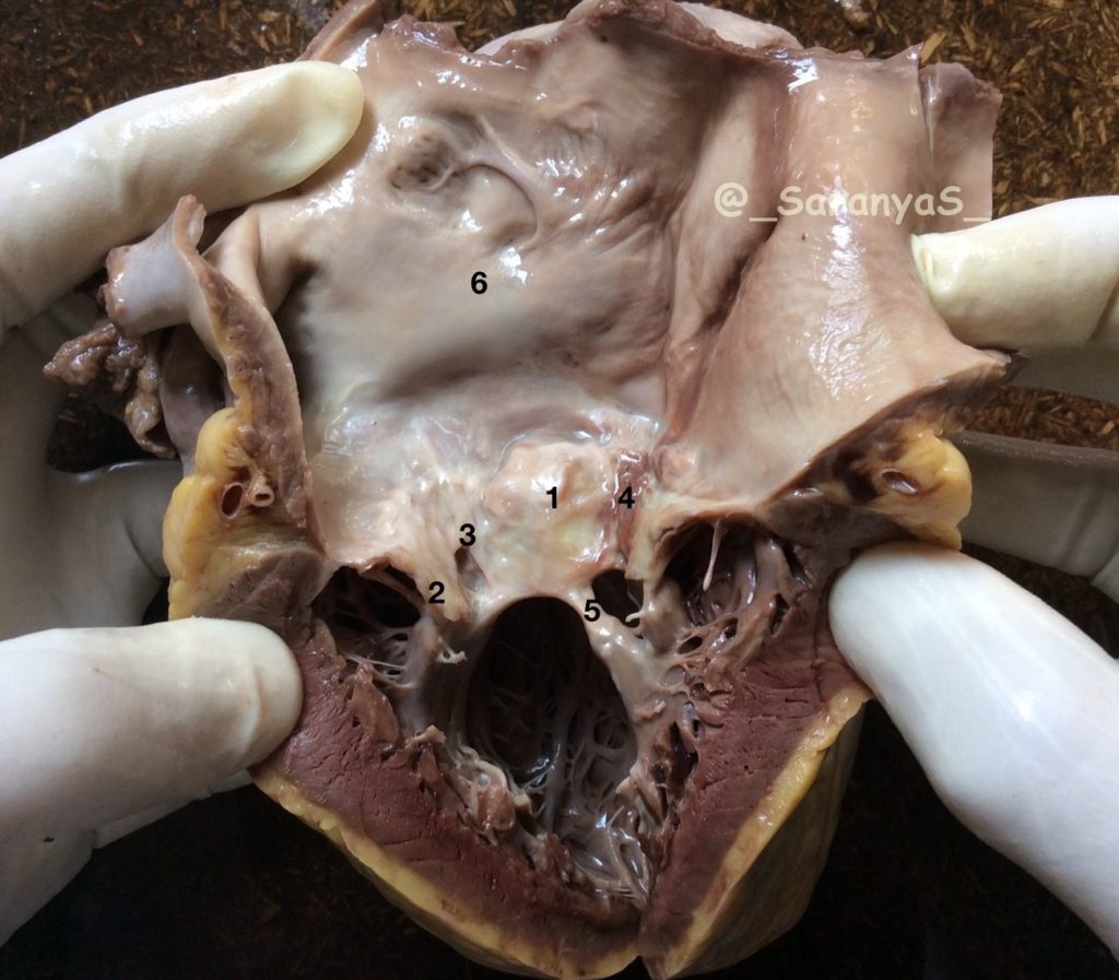
1 – The anterior mitral leaflet is thickened
2 – The posterior mitral leaflet which has been cut while opening the heart is also thickened
3 & 4 – The commissures are fused
5 – The chordae are shortened, thickened and fused
6 – The left atrium is enlarged


Recent Comments