
1. What is cerebrospinal fluid and what are its functions?
CSF is a major fluid in the body. It is produced by the choroid plexus of the two lateral ventricles.
It provides nutrients to the nervous tissue, removes metabolic waste and acts as protective barrier to prevent trauma of the brain and the spinal cord.
It provides nutrients to the nervous tissue, removes metabolic waste and acts as protective barrier to prevent trauma of the brain and the spinal cord.
2. What is the normal volume of CSF?
a. Adults: 90-150 ml
b. Neonates :10-60 ml
b. Neonates :10-60 ml
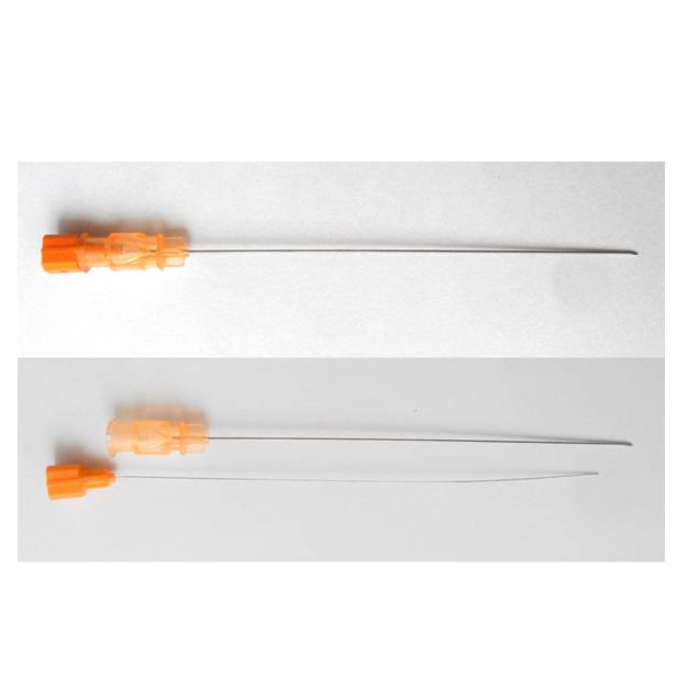
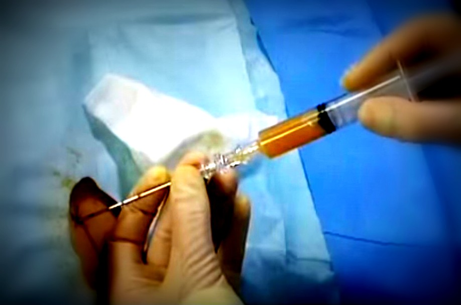
3. How is CSF collected?
It is collected by using lumbar puncture needle inserted at the level of L3/4 or L4/5 interspace..
4. What are the precautions to be taken while collecting the CSF?
The material has to be withdrawn slowly specially during elevated CSF pressure, it requires monitoring of the pressure.
5. In how many tubes cerebrospinal should be collected?
The fluid has to be ideally collected in three tubes and collected in the order in which it is withdrawn.
a. 1st container: for chemical and serological tests
b. 2nd container: for microbiologic examination
c. 3rd container : for cell count.
a. 1st container: for chemical and serological tests
b. 2nd container: for microbiologic examination
c. 3rd container : for cell count.
6. What is the significance of CSF appearance after collection?
The appearance of the fluid immediately after collection gives valuable information listed as below
a. Clear: indicates normal
b. Turbid : indicates inflammatory process like meningitis
c. Bloody: may be due to hemorrhage or traumatic tap
d. Xanthochromic: may indicate old hemorrhage or elevated Bilirubin levels or sometimes even the presence of increased proteins
e. Clotted : indicates clotted blood or any disorder of blood brain barrier where fibrinogen is introduced into the fluid.
7. How do you differentiate between cerebral hemorrhage and traumatic tap when the fluid is bloody?
If it is cerebral hemorrhage, the blood will be evenly distributed in all the three containers, whereas if it is traumatic tap, it is concentrated more in the first container and reduces in subsequent containers.
8. How is cell count done in a Cerebrospinal fluid?
It is done by using a Neubaur counting chamber with a diluted or undiluted fluid. If it is a hemorrhagic fluid, then the diluting fluid will be 3 % glacial acetic acid.
9. How to do a differential cell count in cerebrospinal fluid?
The cerebrospinal fluid should be centrifuged and smears are prepared. The differential count is done on a stained smear.
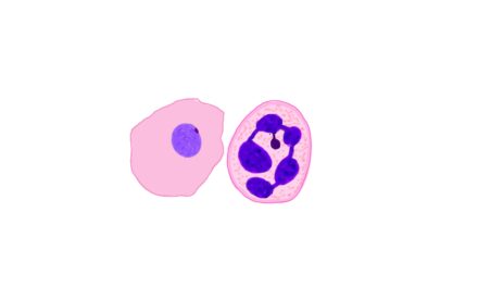
10. What are the methods of estimation of specific gravity of u What are the normal cells found in cerebrospinal fluid.?
Lymphocytes and Monocytes are the normal cells found in cerebrospinal fluid.adults have predominantly lymphocytes where as children have predominantly Monocytes..
11. What is pleocytosis?
Increase in the number of normal cells in the cerebrospinal fluid is called pleocytosis.
12. What are the significance of different cells in cerebrospinal fluid.?
a. Lymphocytes: are seen in normal as well as tubercular and viral meningitis
b. Neutrophils: predominantly in bacterial meningitis. Also can be seen early stages of other meningitis. Can be seen in hemorrhagic tap
c. Monocytes: are normally found as well as in viral and tubercular meningitis
d. Macrophages : indicate old hemorrhage
e. Abnormal cells like blasts are seen in acute leukemias and malignant cells in various primary and metastatic neoplasms..
13. What What is the normal level of protein in cerebrospinal fluid?
The normal levels of protein in cerebrospinal fluid is 15-45mg/dl
14. What are the causes of increased or decreased cerebrospinal fluid protein levels?
a. Increased protein: seen in meningitis, hemorrhage, diabetes, neurosyphilis
b. Decreased protein levels: can be due to rapid production of cerebrospinal fluid or water intoxication.
b. Decreased protein levels: can be due to rapid production of cerebrospinal fluid or water intoxication.
15. What is the normal levels of glucose in cerebrospinal fluid?
The normal levels of glucose is 60 to 70% of the plasma glucose levels.
16. What are the causes of increased and decreased cerebrospinal fluid glucose levels?
a. Increases when the plasma glucose levels increases
b. Decreases in bacterial meningitis..
b. Decreases in bacterial meningitis..
17. What is the role of microbiology tests in cerebrospinal fluid analysis?
The role is identification of causative agents in meningitis. This is usually done by CSF culture.
18. What is the importance of estimation of CSF glutamine level?
Increased levels of glutamine is usually associated with some disturbance in consciousness. Usually done in patients with coma of unknown cause. It is also increased in children with Reye syndrome.
19. What is the role of serologic testing of cerebrospinal fluid.?
The serologic tests are performed to detect the presence of neurosyphilis. VDRL test for syphylis is usually done.
20. What are the laboratory findings in different types of meningitis ?
| Bacterial | Viral | Tubercular | Fungal | |
| Appeareance | Cloudy or purulent | Clear | Clear or opaque. Cob web may be formed | Clear or slightly cloudy |
| Pressure | Elevated | Normal or elevated | Normal or elevated | elevated |
| WBC count | Elevated >1000/microliter | Elevated
10-100/microliter |
Elevated
10-1000/microliter |
Elevated
10-1000/microliter |
| Type of WBC | Neutrophils | Neutrophils in early stages.
Lymphocytes later |
Lymphocytes and monocytes | Lymphocytes and monocytes |
| Proteins | Markedly increases | Moderately increases | Marked | Marked |
| Glucose | Markedly reduced | Normal | Mildly reduced | Normal to reduced |
| Culture | Positive for bacteria | Negative | Positive for tuberculosis | Fungal positive |


Recent Comments