
What are the Organs Involved in Amyloidosis?
The organs involved are as under
Primary Amyloidosis: Heart, GIT, Respiratory tract, peripheral nerves, skin and tongue
Secondary Amyloidosis: Kidney, liver, spleen, lymphnode adrenals and thyroid
Familial Mediterranean fever: Widespread. Kidney, blood vessels , spleen, respiratory tract and liver
Discuss the gross morphology of Amyloidosis
May or may not be visible
If the deposits are too much, then the organ is enlarged, gray, waxy and firm.
How do you demonstrate amyloid in gross specimens
Oldest method, since the time of Virchow! Apply Lugol’s iodine on the cut surface. The area containing amyloid stains deep brown which on Application of dilute sulphuric acid turns blue
The above property is similar to staining property of starch! Hence the name Amyloid!!!
What are the Microscopic features of Amyloid
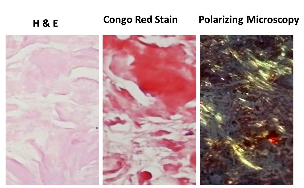
In Hematoxylin & Eosin stain the amyloid,
Is predominantly Extracellular
Can be adjacent to basement membranes, if the deposits are more, it encroaches on cells and destroy
Can be perivascular or vascular
Amorphous, eosinophilic, glassy/hyaline like and Should be differentiated from collagen, fibrin etc
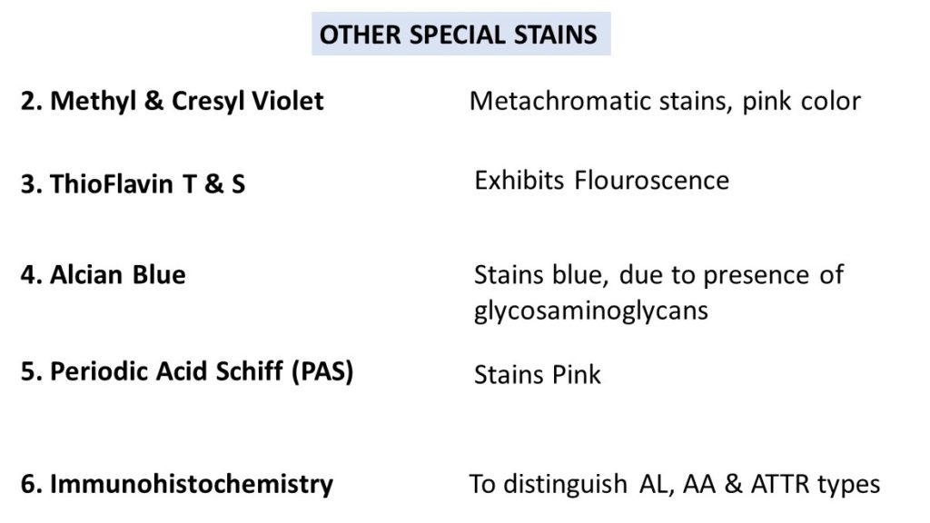
What are the SPECIAL STAINS used to demonstrate amyloid?
- Congo Red Stain : under ordinary light , the amyloid appears red or pink, which when observed under polarizing microscope, show characteristic Apple Green Birefringence. Cross beta pleated sheet confirmation is the reason for this special staining property!
Other special stains as below
AMYLOIDOSIS OF DIFFERENT ORGANS:
Discuss Renal Amyloidosis
- Most common
- Most serious
- Found most commonly in secondary amyloidosis .
- Accounts for one third cases of primary amyloidosis
Gross examination:
- Can be normal size/ enlarged
- may be contracted/shrunken ( due to the narrowing of vessels resulting in ischemia)
- Cut surface: Pale and waxy/ transluscent
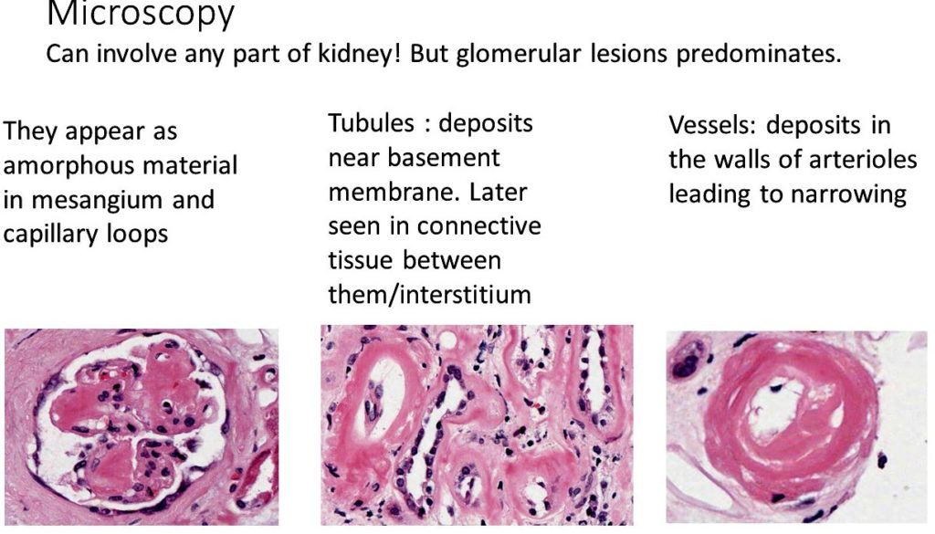
Microscopy as below:
Clinical features of Renal amyloidosis:
- Can present with proteinuria.
- May be severe enough to result in Nephrotic syndrome
- Increased deposition in glomeruli and interstitium may lead to Glomerular ischemia and atrophy of tubules resulting in Chronic Renal failure
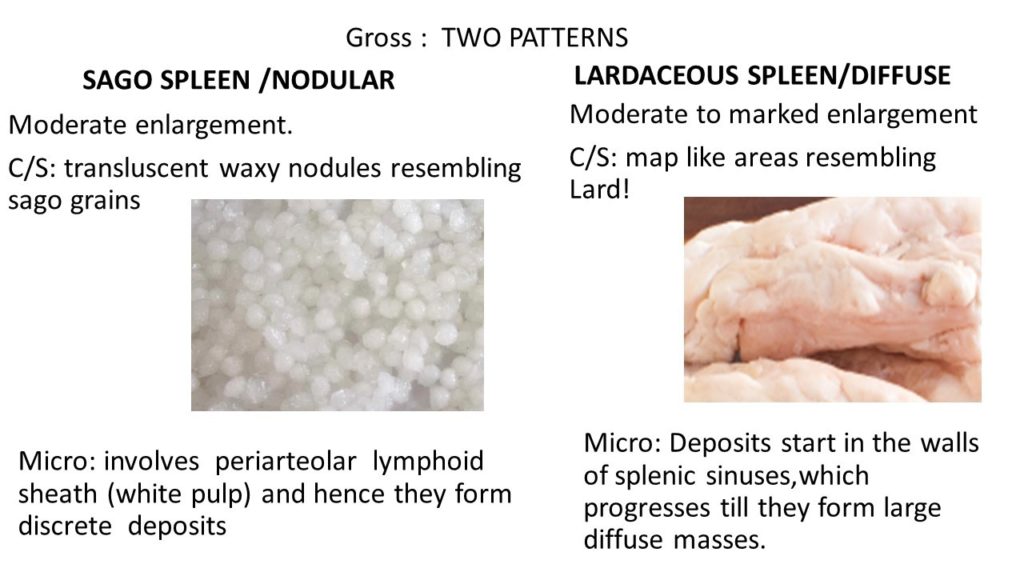
Discuss the features of amyloidosis of Spleen?
The Amyloidosis of spleen is best described in the illustration below
Write briefly on Cardiac amyloidosis:
- Commonly associated with systemic amyloidosis
- Rarely as localized type ( Senile Cardiac Amyloidosis)
Gross :
- May be enlarged.
- Pale , translucent and waxy.
- Nodules/Plaques of amyloid can be seen in the epi/endocardium
Micro:
- Seen in the coronary vessels or surrounding them.
- Around the myocardial fibes ( Ring Fibers)
- Left atrium/interatrial septum in Senile Cardiac Amyloidosis.
Clinical Features:
- Can result in Cardiac Failure.
- Subendocardial deposition can interfere with conduction system leading to arrhythmias .
Discuss the Diagnosis of amyloidosis?
- Despite strong suspicion, the diagnosis of systemic amyloidosis has to be confirmed by tissue diagnosis/ histopathological examination
- Abdominal fat pad aspiration /biopsy is the preferred method for diagnosis because of its Simplicity, low cost, less complications and good accuracy
- Rectal and Gingival biopsy ( previously these were preferred) or labial salivary gland biopsy.
- Localised amyloiadosis: biopsy of the involved tissue and confirmation by staining.
- To know the type of Amyloid Immunohistochemistry is currently the standard method for amyloid typing in routine practice.
What is the Prognosis of Amyloidosis?
- Generalised /systemic amyloidosis: the prognosis is poor.
- Myeloma associated amyloidosis has poorer outcomes.
- localized amyloidosis:local treatment such as excision or laser removal . Recurrence is common
CLICK HERE to watch the video tutorial of Amyloidosis of Part 3


Recent Comments